J Cardiovasc Ultrasound.
2014 Sep;22(3):148-150. 10.4250/jcu.2014.22.3.148.
Native Aortic Valve Thrombosis Resembling Papillary Fibroelastoma
- Affiliations
-
- 1Regional Cardiocerebrovascular Center, Dong-A University Hospital, Busan, Korea. cardiopark@gmail.com
- 2Division of Cardiology, Department of Internal Medicine, Dong-A University College of Medicine, Busan, Korea.
- KMID: 2045432
- DOI: http://doi.org/10.4250/jcu.2014.22.3.148
Abstract
- The differential diagnosis of cardiac mass is important in determining the therapeutic plan and avoiding unnecessary surgical intervention. Non-invasive imaging methods would be useful in the diagnosis of suspected cardiac mass, because they may provide earlier diagnosis and more accurate assessment of cardiac mass. Native aortic valve thrombosis is a rare disorder and difficult to differentiate from a tumor, and in particular, a papillary fibroelastoma. Thus, the clinical decision making with imaging modalities should be performed cautiously. We recently met a female patient who had a aortic valve mass resembling papillary fibroelastoma in normal native valve. The patient underwent a surgical resection and the pathologic finding showed an organized thrombus with no evidence of papillary fibroelastoma.
Keyword
MeSH Terms
Figure
-
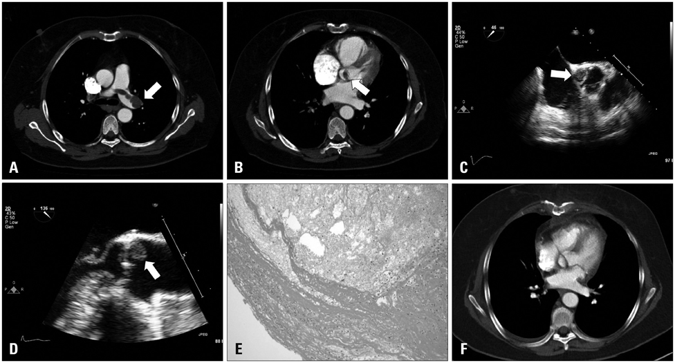
Fig. 1 A: Chest computed tomography with contrast showing pulmonary embolism at the bifurcation of the common pulmonary artery and in its main branches (arrow). B: Computed tomography showing a filling defect attached to aortic valve (arrow). C: Short-axis view of transesophageal echocardiogram showing mass filling the non-coronary sinus of Valsalva (arrow). D: Long-axis view of transesophageal echocardiogram showing a mass just behind the non-coronary cusp (arrow). E: Histologic photomicrography of cardiac mass showing organized thrombus (hematoxylin & eosin stain, × 40). F: Follow-up computed tomography after surgical excision showing complete disappearance of the thrombus.
Reference
-
1. Sun JP, Asher CR, Yang XS, Cheng GG, Scalia GM, Massed AG, Griffin BP, Ratliff NB, Stewart WJ, Thomas JD. Clinical and echocardiographic characteristics of papillary fibroelastomas: a retrospective and prospective study in 162 patients. Circulation. 2001; 103:2687–2693.
Article2. Yamawaki M, Shimoyama M, Furuse Y, Kinugasa Y, Ogino K, Enokida M, Teshima R, Kitamura Y, Hayashi K, Shigemasa C. Floating thrombus arising from left sinus of Valsalva induced intermittent occlusion of left coronary artery and caused cardiogenic shock. Int J Cardiol. 2007; 114:272–273.
Article3. Nagata Y, Miyamoto T, Komura M, Niwa A, Kawaguchi S, Shirai T, Fujiwara H, Isobe M. Giant organized thrombus in the left sinus of valsalva causing intermittent left coronary obstruction: an unusual case of acute myocardial infarction. Circ J. 2004; 68:795–798.
Article4. Cho SJ, Yang JH, Shin JU, Uhm JE, Lee SC, Park SW, Park PW. A case of spontaneous native aortic valvular thrombosis that caused aortic stenoinsufficiency in the bicuspid aortic valve. Korean Circ J. 2006; 36:666–668.
Article5. Grondin F, Giannoccaro JP. Antiphospholipid antibody syndrome associated with large aortic valve vegetation and stroke. Can J Cardiol. 1995; 11:133–135.6. Hegde VA, Vivas Y, Shah H, Haybron D, Srinivasan V, Dua A, Gradman A. Cardiovascular surgical outcomes in patients with the antiphospholipid syndrome--a case-series. Heart Lung Circ. 2007; 16:423–427.
Article7. Weiss S, Nyzio JB, Cines D, Detre J, Milas BL, Narula N, Floyd TF. Antiphospholipid syndrome: intraoperative and postoperative anticoagulation in cardiac surgery. J Cardiothorac Vasc Anesth. 2008; 22:735–739.
Article8. Djokovic A, Stojanovich L, Kontic M, Stanisavljevic N, Radovanovic S, Marisavljevic D. Association between cardiac manifestations and antiphospholipid antibody type and level in a cohort of Serbian patients with primary and secondary antiphospholipid syndrome. Isr Med Assoc J. 2014; 16:162–167.9. Rahbar K, Seifarth H, Schäfers M, Stegger L, Hoffmeier A, Spieker T, Tiemann K, Maintz D, Scheld HH, Schober O, Weckesser M. Differentiation of malignant and benign cardiac tumors using 18F-FDG PET/CT. J Nucl Med. 2012; 53:856–863.
Article10. Kim AY, Kim JS, Yoon Y, Kim EJ. Multidetector computed tomography findings of a papillary fibroelastoma of the aortic valve: a case report. J Korean Med Sci. 2010; 25:809–812.
Article
- Full Text Links
-
- Actions
-
Cited
- CITED
-
- Close
- Share
-
- Similar articles
-
- Aortic Valve Papillary Fibroelastoma Triggering Chest Pain: A case report
- Aortic Valve Papillary Fibroelastoma: Report of 1 Case
- Papillary Fibroelastoma of the Aortic Valve: Discovered by Chance with Intraoperative Transesophageal Echocardiography: A case report
- Multiple Cardiac Papillary Fibroelastoma of the Aortic Valve
- Multidetector Computed Tomography Findings of a Papillary Fibroelastoma of the Aortic Valve: A Case Report
