Multidetector Computed Tomography Findings of a Papillary Fibroelastoma of the Aortic Valve: A Case Report
- Affiliations
-
- 1Department of Radiology, Dongguk University Ilsan Hospital, Dongguk University, Goyang, Korea. kjs7143@kornet.net
- 2Department of Thoracic and Cardiovascular Surgery, Dongguk University Ilsan Hospital, Dongguk University, Goyang, Korea.
- KMID: 1713972
- DOI: http://doi.org/10.3346/jkms.2010.25.5.809
Abstract
- Papillary fibroelastoma is a rare benign cardiac tumor that represents 10% of all primary cardiac tumors. Diagnosis is accomplished incidentally by echocardiography that is usually performed for another purpose. Most papillary fibroelastomas are asymptomatic, but the lesions are recognized as a cause of embolisms. To the best of our knowledge, there has been no case report of computed tomography findings of a papillary fibroelastoma. We report a case of a papillary fibroelastoma in a 78-yr-old woman who had dyspnea and chest tightness. Echocardiography revealed a small lobulated mobile echogenic mass attached to the aortic valve, and CT demonstrated a lobulated soft tissue density mass with a thin stalk at the sinotubular junction of the aortic valve.
MeSH Terms
Figure
-
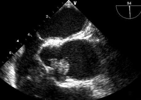
Fig. 1 Transesophageal echocardiography shows a 15×10 mm sized lobulated echogenic mass attached to the sinotubular junction of the aortic valve.
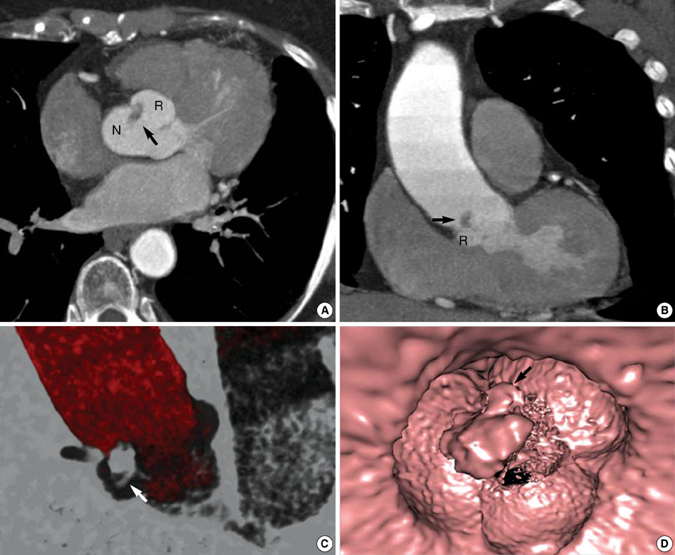
Fig. 2 Axial (A) and coronal (B) CT images show an 11 mm lobulated soft tissue density mass (arrow) at the sinotubular junction between the right coronary cusp (R) and noncoronary cusp (N) of the aortic valve. A volume-rendered image (C) shows a thin stalk (arrow) that is connected between the lesion and the aortic valve. A virtual aortoscopic image (D) shows a lobulated mass (arrow).

Fig. 3 A gross specimen demonstrates the fibroelastoma appearing as an approximate 11 mm sized lesion with multiple threads on the surface and some gelatinous materials in part of the mass.
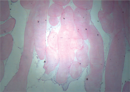
Fig. 4 A photomicrograph shows that the papillary fronds consist of dense connective tissue core surrounded by flat endothelial cells (H&E; original magnification,×10).
Cited by 1 articles
-
Native Aortic Valve Thrombosis Resembling Papillary Fibroelastoma
Minkwan Kim, Suk-Hyun Kim, Sang Yi Moon, Eu Gene Jeong, Eui Han Jung, Hwa Seong Nam, Jae-Hyuk Choi, Kyungil Park
J Cardiovasc Ultrasound. 2014;22(3):148-150. doi: 10.4250/jcu.2014.22.3.148.
Reference
-
1. Tazelaar HD, Locke TJ, McGregor CG. Pathology of surgically excised primary cardiac tumors. Mayo Clin Proc. 1992. 67:957–965.
Article2. Burke A, Virmani R. Tumors of the heart and great vessels. Atlas of tumor pathology: fasc 16, ser 3. 1996. Washington, DC: Armed Forces Institute of Pathology;1–98.3. Edwards FH, Hale D, Cohen A, Thompson L, Pezzella AT, Virmani R. Primary cardiac valve tumors. Ann Thorac Surg. 1991. 52:1127–1131.
Article4. Grebenc ML, Rosado de Christenson ML, Burke AP, Green CE, Galvin JR. Primary cardiac and pericardial neoplasms: radiologicpathologic correlation. Radiographics. 2000. 20:1073–1103.
Article5. Sparrow PJ, Kurian JB, Jones TR, Sivananthan MU. MR imaging of cardiac tumors. Radiographics. 2005. 25:1255–1276.
Article6. Wintersperger BJ, Becker CR, Gulbins H, Knez A, Bruening R, Heuck A, Reiser MF. Tumors of the cardiac valves; imaging findings in magnetic resonance imaging, electron beam computed tomography and echocardiography. Eur Radiol. 2000. 10:443–449.
Article7. Shiraishi J, Tagawa M, Yamada T, Sawada T, Tatsumi T, Azuma A, Shimada Y, Yaku H, Kitamura N, Nakagawa M. Papillary fibroelastoma of the aortic valve: evaluation with transoesophageal echocardiography and magnetic resonance imaging. Jpn Heart J. 2003. 44:799–803.8. Araoz PA, Mulvagh SL, Tazelaar HD, Julsrud PR, Breen JF. CT and MR imaging of benign primary cardiac neoplasms with echocardiographic correlation. Radiographics. 2000. 20:1303–1319.
Article9. Ramesh MG, Ijaz AK, Chandra KN, Nirav JM, Balendu CV, Terrence JS. Cardiac papillary fibroelastoma: A comprehensive analysis of 725 cases. Am Heart J. 2003. 146:404–410.10. Grinda JM, Couteil JP, Chauvaud S, D'Attellis N, Berrebi A, Fabiani JN, Deloche A, Carpentier A. Cardiac valve papillary fibroelastoma: surgical excision for revealed or potential embolization. J Thorac Cardiovasc Surg. 1999. 117:106–110.
Article11. Frederick JS. Ramzi SC, Vinay K, Tucker C, editors. The Heart. Robbins pathologic basis of disease. 1999. 6th ed. USA: W.B. Sanders Company;590–591.12. Klarich KW, Enriquez-Sarano M, Gura GM, Edwards WD, Tajik AJ, Seward JB. Papillary fibroelastoma: Echocardiographic Characteristics for Diagnosis and Pathologic Correlation. J Am Coll Cardiol. 1997. 30:784–790.
Article13. al-Mohammad A, Pambakian H, Young C. Fibroelastoma: case report and review of the literature. Heart. 1998. 79:301–304.
Article
- Full Text Links
-
- Actions
-
Cited
- CITED
-
- Close
- Share
-
- Similar articles
-
- Aortic Valve Papillary Fibroelastoma Triggering Chest Pain: A case report
- Aortic Valve Papillary Fibroelastoma: Report of 1 Case
- Papillary Fibroelastoma of the Aortic Valve: Discovered by Chance with Intraoperative Transesophageal Echocardiography: A case report
- Native Aortic Valve Thrombosis Resembling Papillary Fibroelastoma
- Multiple Cardiac Papillary Fibroelastoma of the Aortic Valve
