Bilateral Vertebral Artery Dissecting Aneurysms Presenting with Subarachnoid Hemorrhage Treated by Staged Coil Trapping and Covered Stents Graft
- Affiliations
-
- 1Department of Neurosurgery, Soonchunhyang University Cheonan Hospital, Cheonan, Korea. smyoon@sch.ac.kr
- 2Department of Neurosurgery, Soonchunhyang University Gumi Hospital, Gumi, Korea.
- 3Department of Neurosurgery, Soonchunhyang University Hospital, Seoul, Korea.
- KMID: 2018190
- DOI: http://doi.org/10.3340/jkns.2012.51.3.155
Abstract
- The treatment of bilateral vertebral artery dissecting aneurysms (VADAs) presenting with subarachnoid hemorrhage (SAH) is still challenging. The authors report a rare case of bilateral VADA treated with coil trapping of ruptured VADA and covered stents implantation after multiple unsuccessful stent assisted coiling of the contralateral unruptured VADA. A 44-year-old woman was admitted to our hospital because of severe headache and sudden stuporous consciousness. Brain CT showed thick SAH and intraventricular hemorrhage. Cerebral angiography demonstrated bilateral VADA. Based on the SAH pattern and aneurysm configurations, the right VADA was considered ruptured. This was trapped with endovascular coils without difficulty. One month later, the contralateral unruptured VADA was protected using a stent-within-a-stent technique, but marked enlargement of the left VADA was detected by 8-months follow-up angiography. Subsequently two times coil packing for pseudosacs resulted in near complete occlusion of left VADA. However, it continued to grow. Covered stents graft below the posterior inferior cerebellar artery (PICA) origin and a coronary stent implantation across the origin of the PICA resulted in near complete obliteration of the VADA. Covered stent graft can be used as a last therapeutic option for the management of VADA, which requires absolute preservation of VA flow.
Keyword
MeSH Terms
Figure
-

Fig. 1 Pre- and post-operative radiologic images in a 44-year-old woman presenting with SAH. Brain CT (A) shows thick SAH on basal, prepontine, and both cerebellopontine angle cisterns, and acute hydrocephalus due to intraventricular hemorrhage. Preoperative digital subtraction angiography (B) and 3-dimensional rotational angiogram (C) demonstrate a 6×12 mm sized right VADA (the right PICA is not visualized), and a 5×7 mm sized left VADA, which is incorporated with PICA origin. Postoperative vertebral angiograms after packing of 19 coils for right VADA show complete trapping of the right VADA with sufficient flow to the basilar artery via the left vertebral artery (D and E). Vertebral angiogram after double stent implantation for the left VADA demonstrates slightly reduced blood flow to the aneurysmal sac (F). SAH : subarachnoid hemorrhage, VADA : vertebral artery dissecting aneurysm, PICA : posterior inferior cerebellar artery.
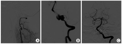
Fig. 2 Follow-up vertebral angiograms at 8 months after double overlapping stenting on the left VADA showing stable occlusion of right VADA (A) and marked growth of the left VADA (B). Postoperative left vertebral angiogram after coil packing into the two different pseudosacs shows near complete obliteration of left VADA, while sparing left PICA flow (C). VADA : vertebral artery dissecting aneurysm, PICA : posterior inferior cerebellar artery.
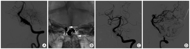
Fig. 3 Follow-up left vertebral angiogram (A) at 5 months after coil packing of the left VADA demonstrating its further growth. Postoperative skull radiograph (B) shows triple overlapping stents and coil mass on the left vertebral artery after further coil packing of the two growing pseudosacs and bailout stenting using an Enterprise stent. White arrows indicate proximal and distal end of the triple stents. Postoperative angiograms (C and D) show near complete obliteration of the VADA and sparing of left PICA flow. VADA : vertebral artery dissecting aneurysm, PICA : posterior inferior cerebellar artery.

Fig. 4 Follow-up angiograms at 4 months after final embolization also showing left VADA regrowth (A). Postoperative skull radiograph after telescopic stenting with two covered stents graft (Graftmaster 3.5×12 mm, 3.5×9 mm) on the left vertebral artery just below the PICA origin and a third stent (Driver 3.5×12 mm) across the PICA origin (B). Arrows indicate the proximal end of covered stent and distal end of Driver stent. Final vertebral angiograms at early arterial (C) and late arterial phase (D) still demonstrate slow retrograde flow to VADA, although flow velocity into the VADA is markedly decreased. Follow-up angiograms 2 month later show near complete occlusion of the left VADA (E and F). VADA : vertebral artery dissecting aneurysm, PICA : posterior inferior cerebellar artery.
Cited by 2 articles
-
The Difference of Each Angiographic Finding After Multiple Stent According to Stent Type in Bilateral Vertebral Artery Dissection
Dong Hoon Lee, Woon Ki Yoon, Min Woo Baik, Hoon Kim
J Cerebrovasc Endovasc Neurosurg. 2013;15(3):229-234. doi: 10.7461/jcen.2013.15.3.229.Are Blood Blister-Like Aneurysms a Specific Type of Dissection? A Comparative Study of Blood Blister-Like Aneurysms and Ruptured Mizutani Type 4 Vertebral Artery Dissections
Sook Young Sim, Joonho Chung, Yong Sam Shin
J Korean Neurosurg Soc. 2014;56(5):395-399. doi: 10.3340/jkns.2014.56.5.395.
Reference
-
1. Ahn JY, Han IB, Kim TG, Yoon PH, Lee YJ, Lee BH, et al. Endovascular treatment of intracranial vertebral artery dissections with stent placement or stent-assisted coiling. AJNR Am J Neuroradiol. 2006; 27:1514–1520. PMID: 16908571.2. Ding H, He M, You C, Deng L. [Application of endovascular covered stent for treating vertebral dissecting aneurysm and carotid-cavernous fistula]. Zhongguo Xiu Fu Chong Jian Wai Ke Za Zhi. 2009; 23:215–218. PMID: 19275107.3. He M, Zhang H, Lei D, Mao BY, You C, Xie XD, et al. Application of covered stent grafts for intracranial vertebral artery dissecting aneurysms. J Neurosurg. 2009; 110:418–426. PMID: 18847343.
Article4. Inoue A, Kohno K, Takechi A, Kohno K, Matsushige T, Takeda T. Bilateral vertebral artery dissecting aneurysm with subarachnoid hemorrhage treated with staged bilateral vertebral artery coil occlusion : a case report. Surg Neurol. 2008; 70:319–322. discussion 322. PMID: 18207505.
Article5. Katsuno M, Mizunari T, Kobayashi S, Takahashi H, Teramoto A. Rupture of a vertebral artery dissecting aneurysm developing immediately after trapping of a dissecting aneurysm on the contralateral vertebral artery : case report. Neurol Med Chir (Tokyo). 2009; 49:468–470. PMID: 19855144.
Article6. Koh JS, Ryu CW, Lee SH, Bang JS, Kim GK. Bilateral vertebral-artery-dissecting aneurysm causing subarachnoid hemorrhage cured by staged endovascular reconstruction after occlusion. Cerebrovasc Dis. 2009; 27:202–204. PMID: 19153480.
Article7. Lieber BB, Stancampiano AP, Wakhloo AK. Alteration of hemodynamics in aneurysm models by stenting : influence of stent porosity. Ann Biomed Eng. 1997; 25:460–469. PMID: 9146801.
Article8. Lylyk P, Cohen JE, Ceratto R, Ferrario A, Miranda C. Combined endovascular treatment of dissecting vertebral artery aneurysms by using stents and coils. J Neurosurg. 2001; 94:427–432. PMID: 11235947.
Article9. Matsumoto K, Akagi K, Abekura M, Sakaguchi T. Obliteration of bilateral dissecting aneurysms of the vertebral arteries following repeated subarachnoid hemorrhage : a case report. Neurol Res. 2002; 24:837–841. PMID: 12500710.
Article10. Nashimoto T, Komata T, Honma J, Yamashita S, Seki Y, Kurashima A, et al. Successful treatment of bilateral vertebral artery dissecting aneurysms with subarachnoid hemorrhage : report of three cases. J Stroke Cerebrovasc Dis. 2010; in press.11. Otawara Y, Ogasawara K, Ogawa A, Kogure T. Dissecting aneurysms of the bilateral vertebral arteries with subarachnoid hemorrhage : report of three cases. Neurosurgery. 2002; 50:1372–1374. discussion 1374-1375. PMID: 12015860.
Article12. Park SI, Kim BM, Kim DI, Shin YS, Suh SH, Chung EC, et al. Clinical and angiographic follow-up of stent-only therapy for acute intracranial vertebrobasilar dissecting aneurysms. AJNR Am J Neuroradiol. 2009; 30:1351–1356. PMID: 19342544.
Article13. Redekop G, Marotta T, Weill A. Treatment of traumatic aneurysms and arteriovenous fistulas of the skull base by using endovascular stents. J Neurosurg. 2001; 95:412–419. PMID: 11565861.
Article14. Redekop G, TerBrugge K, Willinsky R. Subarachnoid hemorrhage from vertebrobasilar dissecting aneurysm treated with staged bilateral vertebral artery occlusion : the importance of early follow-up angiography : technical case report. Neurosurgery. 1999; 45:1258–1262. discussion 1262-1263. PMID: 10549948.15. Sakamoto S, Ohba S, Shibukawa M, Kiura Y, Okazaki T, Arita K, et al. Staged bilateral vertebral artery occlusion for ruptured dissecting aneurysms of the basilar artery : a report of 2 cases. Surg Neurol. 2005; 64:456–461. discussion 461. PMID: 16253701.
Article16. Somekawa K, Nagata K, Kawamoto S, Furuya H, Tanioka D, Isoo A. [Treatment for the ruptured bilateral vertebral dissecting aneurysms]. No Shinkei Geka. 2002; 30:321–325. PMID: 11905026.17. Stone GW, Goldberg S, O'Shaughnessy C, Midei M, Siegel RM, Cristea E, et al. 5-year follow-up of polytetrafluoroethylene-covered stents compared with bare-metal stents in aortocoronary saphenous vein grafts the randomized (BARRICADE barrier approach to restenosis : restrict intima to curtail adverse events) trial. JACC Cardiovasc Interv. 2011; 4:300–309. PMID: 21435608.
Article18. Suh SH, Kim BM, Park SI, Kim DI, Shin YS, Kim EJ, et al. Stent-assisted coil embolization followed by a stent-within-a-stent technique for ruptured dissecting aneurysms of the intracranial vertebrobasilar artery. Clinical article. J Neurosurg. 2009; 111:48–52. PMID: 19326976.
Article19. Yoon WK, Kim YW, Kim SR, Park IS, Kim SD, Jo KW, et al. Angiographic and clinical outcomes of stent-alone treatment for spontaneous vertebrobasilar dissecting aneurysm. Acta Neurochir (Wien). 2010; 152:1477–1486. discussion 1486. PMID: 20508955.
Article20. Zenteno MA, Santos-Franco JA, Freitas-Modenesi JM, Gómez C, Murillo-Bonilla L, Aburto-Murrieta Y, et al. Use of the sole stenting technique for the management of aneurysms in the posterior circulation in a prospective series of 20 patients. J Neurosurg. 2008; 108:1104–1118. PMID: 18518712.
Article
- Full Text Links
-
- Actions
-
Cited
- CITED
-
- Close
- Share
-
- Similar articles
-
- Bilateral Vertebral Artery Dissecting Aneurysms: A Long Term Follow-up Results of Microsurgical Trapping and Proximal Occlusion
- Endovascular Coil Trapping of a Ruptured Dissecting Aneurysm of the Vertebral Artery Using Detachable Coils and Micro-Tornado(R) Coils
- Early Rebleeding after Internal Trapping of a Ruptured Vertebral Artery Dissecting Aneurysm: A Case Report
- Cerebral Bypass Surgery for Treating Unclippable and Uncoilable Aneurysms
- Dissecting Aneurysm Associated with a Double Origin of the Posterior Inferior Cerebellar Artery Causing Subarachnoid Hemorrhage
