Dissecting Aneurysm Associated with a Double Origin of the Posterior Inferior Cerebellar Artery Causing Subarachnoid Hemorrhage
- Affiliations
-
- 1Department of Neurosurgery, Kyung Hee University Hospital at Gangdong, Seoul, Korea. neurokoh@hanmail.net
- KMID: 2190468
- DOI: http://doi.org/10.3340/jkns.2012.51.1.40
Abstract
- Two cases of the posterior fossa dissecting aneurysm associated with a double origin of the posterior inferior cerebellar artery (DOPICA) causing subarachnoid hemorrhage are presented. After observing a relationship between the aneurysm and DOPICA on a three dimensional rotational angiogram (3DRA), the dissecting aneurysms were successfully obliterated by surgical trapping and endovascular internal trapping, respectively. This report warrants suspecting DOPICA of an associating anomaly predisposing to dissecting aneurysm in the vertebral artery-posterior inferior cerebellar artery territory and highlights the role of 3DRA in pretreatment evaluation of unusual aneurysms accompanying a particular anatomical variation.
Keyword
Figure
-
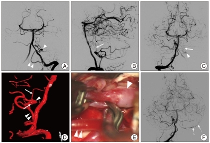
Fig. 1 Case 1. Anteroposterior (AP) view of left vertebral artery (VA) angiogram (A) showing a peripheral aneurysm (arrow head) located on the hemispheric branch of the posterior inferior cerebellar artery (PICA) which has a low origin on the V4 (double arrowhead). Lateral view of left VA angiogram (B) demonstrating that a small cranial branch (arrow) originating from the distal V4 is directly anastomosed with the PICA. AP view of right VA angiogram (C) showing contrast filling of the aneurysm (arrowhead) through a small branch (arrow) originating on the distal V4. Reconstructed 3-dimensional rotational angiogram (D) clearly demonstrating the upper (arrows) and lower (double arrowheads) channel of double origin of the PICA and an associating aneurysm (arrowhead). Intraoperative photograph (E) showing trapping of the distal PICA (double arrowheads) harboring aneurysm (arrowhead). Follow-up right VA angiogram obtained 3 weeks after surgical trapping (F) showing a patent flow in the hemispheric branch of the left PICA (broken arrows) by collaterals and complete exclusion of the dissecting aneurysm.
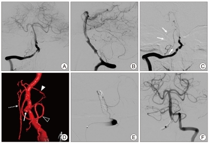
Fig. 2 Case 2. Right vertebral artery (VA) angiogram (A and B) obtained at admission showing an irregular constriction and outpouching of the right VA and revealing an extracranial origin PICA. Follow-up right VA angiogram obtained 3 days after admission (C) showing growing of aneurismal dilations from which a faint vessel arises (arrows). Enlarged, oblique view, 3DRA of the right VA (D) demonstrating that a small upper branch (arrowhead) originating from the proximal aneurysm (arrow) is directly anastomosed with the PICA (open arrowhead). The anterior spinal artery (broken arrow) originating on the V4 is located just distal to the dissection. Postoperative, lateral view of right VA angiogram (E) and AP view of left VA angiogram (F) showing total occlusion of the right VA in which are detachable coils (asterisk). PICA : posterior inferior cerebellar artery, 3DRA : three dimensional rotational angiogram, AP : anteroposterior.
Cited by 1 articles
-
In Situ Intersegmental Anastomosis within a Single Artery for Treatment of an Aneurysm at the Posterior Inferior Cerebellar Artery: Closing Omega Bypass
Sung Ho Lee, Seok Keun Choi
J Korean Neurosurg Soc. 2015;58(5):467-470. doi: 10.3340/jkns.2015.58.5.467.
Reference
-
1. Ali MJ, Bendok BR, Tawk RG, Getch CC, Batjer HH. Trapping and revascularization for a dissecting aneurysm of the proximal posteroinferior cerebellar artery : technical case report and review of the literature. Neurosurgery. 2002; 51:258–262. discussion 262-263. PMID: 12182429.2. Arai K, Endo S, Hirashima Y, Takaku A. Posterior inferior cerebellar artery aneurysm associated with fenestration of the vertebral artery--case report. Neuro Med Chir (Tokyo). 1989; 29:29–31.
Article3. Ashley WW Jr, Chicoine MR. Subarachnoid hemorrhage caused by posterior inferior cerebellar artery aneurysm with an anomalous course of the atlantoaxial segment of the vertebral artery. Case report and review of literature. J Neurosurg. 2005; 103:356–360. PMID: 16175868.
Article4. Fine AD, Cardoso A, Rhoton AL Jr. Microsurgical anatomy of the extracranial-extradural origin of the posterior inferior cerebellar artery. J Neurosurg. 1999; 91:645–652. PMID: 10507387.
Article5. Kwon BJ, Jung C, Im SH, Lee DH, Han MH. Double origin of the posteroinferior cerebellar artery : angiographic anatomy and endovascular treatment of concurrent vertebrobasilar dissection. Neurosurgery. 2007; 61:242–247. discussion 247-248. PMID: 18091238.6. Lasjaunias P, Vallee B, Person H, Ter Brugge K, Chiu M. The lateral spinal artery of the upper cervical spinal cord. Anatomy, normal variations, and angiographic aspects. J Neurosurg. 1985; 63:235–241. PMID: 4020445.7. Lesley WS, Dalsania HJ. Double origin of the posterior inferior cerebellar artery. AJNR Am J Neuroradiol. 2004; 25:425–427. PMID: 15037467.8. Lesley WS, Rajab MH, Case RS. Double origin of the posterior inferior cerebellar artery : association with intracranial aneurysm on catheter angiography. AJR Am J Roentgenol. 2007; 189:893–897. PMID: 17885063.
Article9. Nishizaki T, Tamaki N, Nishida Y, Fujita K, Matsumoto S. Aneurysms of the distal posterior inferior cerebellar artery : experience with three cases and review of the literature. Neurosurgery. 1985; 16:829–832. PMID: 4010907.
Article10. Pasco A, Thouveny F, Papon X, Tanguy JY, Mercier P, Caron-Poitreau C, et al. Ruptured aneurysm on a double origin of the posterior inferior cerebellar artery : a pathological entity in an anatomical variation. Report of two cases and review of the literature. J Neurosurg. 2002; 96:127–131. PMID: 11794593.
Article11. Tabatabai SA, Zadeh MZ, Meybodi AT, Hashemi M. Extracranial aneurysm of the posterior inferior cerebellar artery with an aberrant origination : case report. Neurosurgery. 2007; 61:E1097–E1098. discussion E1098. PMID: 18091258.
- Full Text Links
-
- Actions
-
Cited
- CITED
-
- Close
- Share
-
- Similar articles
-
- The Dissecting Aneurysm of the Posterior Inferior Cerebellar Artery with Unusual Clinical Course
- Stent-Jack Technique for Ruptured Vertebral Artery Dissecting Aneurysm Involving the Origin of Posterior Inferior Cerebellar Artery
- In Situ Intersegmental Anastomosis within a Single Artery for Treatment of an Aneurysm at the Posterior Inferior Cerebellar Artery: Closing Omega Bypass
- Aneurysm of Distal Posterior Inferior Cerebellar Artery:Case Report with Review of the Literature
- Cerebral Dissecting Aneurysms in Patients with Essential Thrombocythemia
