Perianal Mucinous Adenocarcinoma with an Emphasis on the MR Imaging Features: A Case Report
- Affiliations
-
- 1Department of Diagnostic Radiology, College of Medicine, Inje University Pusan Paik Hospital, Korea. nayaa_neo@naver.com
- 2Department of Pathology, Changwon Fatima Hospital, Korea.
- 3Department of General Surgery, Changwon Fatima Hospital, Korea.
- 4Department of Diagnostic Radiology, Changwon Fatima Hospital, Korea.
- KMID: 2002956
- DOI: http://doi.org/10.3348/jksr.2010.62.5.471
Abstract
- An 80-year-old man, who presented with a perianal mass, showed a multilocular mass with peripheral calcification in the retroanal region at CT. The MR imaging detected a mass invading into the posterior aspect of the external anal sphincter, and was shown as having a high T1 and T2 signal intensity with a different T1 signal intensity in each locule. After contrast injection, septal and peripheral enhancement of the tumor was observed. Surgery was performed and revealed a perianal mucinous adenocarcinoma. To the best of our knowledge, this is the first report describing the MR features of a perianal mucinous adenocarcinoma in the Korean literature. We described a case of perianal mucinous adenocarcinoma with an emphasis on the MR imaging features.
MeSH Terms
Figure
-
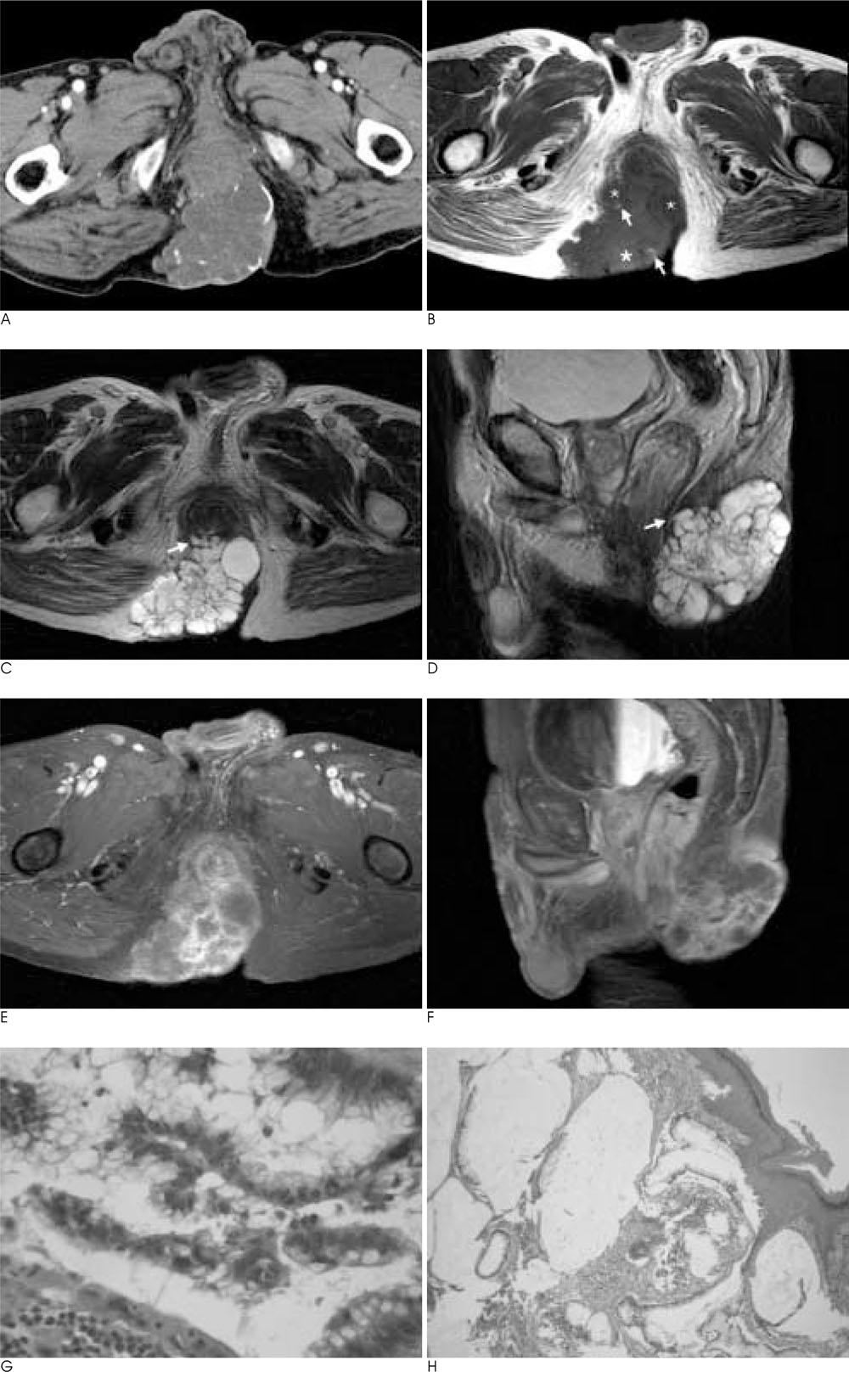
Fig. 1 An 80-year-old man with a surgically proven perianal mucinous adenocarcinoma. A. An axial CT image after administration of contrast material shows the presence of a multilocular mass with multiple marginal calcifications. The mass is attached to the posterior aspect of the anus and extends to the skin. B. T1-weighted MR image shows a slightly high signal intensity comparing to that of muscle, with a different internal signal intensity for each locule (small *: relatively hyposignal intensity, large *: relatively hypersignal intensity). Curvilinear high signal intensities along the margin (arrows) of some locules, which are consistent with the marginal calcification on CT, are also identified. C, D. On the axial and sagittal T2 weighted MR images, the multilocular mass is shown as having a markedly high signal intensity and a focal invasion into the posterior aspect of the extrinsic anal sphincter is identified (arrows). E, F. Axial and sagittal fat-saturated gadolinium-enhanced T1 weighted MR images show septal and peripheral enhancement of the tumor. G. The multilocular cystic wall is lined by mucinous columnar cells. An overlying perianal squamous epithelial layer is seen (H & E staining, × 40). H. The glandular epithelial cells have hyperchromatic and atypical nuclei with nuclear stratification (H & E staining, × 400).
Reference
-
1. Wong AY, Rahilly MA, Adams W, Lee CS. Mucinous anal gland carcinoma with perianal pagetoid spread. Pathology. 1998; 30:1–3.2. Abel ME, Chiu YS, Russell TR, Volpe PA. Adenocarcinoma of the anal glands. Results of a survey. Dis Colon Rectum. 1993; 36:383–387.3. Nishimura T, Nozue M, Suzuki K, Imai M, Suzuki S, Sakahara H, et al. Perianal mucinous carcinoma successfully treated with a combination of external beam radiotherapy and high dose rate interstitial brachytherapy. Br J Radiol. 2000; 73:661–664.4. Yang DM, Jung DH, Kim H, Kang JH, Kim SH, Kim JH, et al. Retroperitoneal cystic masses: CT, clinical, and pathologic findings and literature review. Radiographics. 2004; 24:1353–1365.5. Hama Y, Makita K, Yamana T, Dodanuki K. Mucinous adenocarcinoma arising from fistula in ano: MRI findings. AJR Am J Roentgenol. 2006; 187:517–521.6. Fujimoto H, Ikeda M, Shimofusa R, Terauchi M, Eguchi M. Mucinous adenocarcinoma arising from fistula-in-ano: findings on MRI. Eur Radiol. 2003; 13:2053–2054.7. Hussain SM, Outwater EK, Siegelman ES. Mucinous versus nonmucinous rectal carcinomas: differentiation with MR imaging. Radiology. 1999; 213:79–85.8. Okamoto Y, Tanaka YO, Tsunoda H, Yoshikawa H, Minami M. Malignant or borderline mucinous cystic neoplasms have a larger number of loculi than mucinous cystadenoma: a retrospective study with MR. J Magn Reson Imaging. 2007; 26:94–99.9. Henkelman RM, Watts JF, Kucharczyk W. High signal intensity in MR images of calcified brain tissue. Radiology. 1991; 179:199–206.10. Anthony T, Simmang C, Lee EL, Turnage RH. Perianal mucinous adenocarcinoma. J Surg Oncol. 1997; 64:218–221.
- Full Text Links
-
- Actions
-
Cited
- CITED
-
- Close
- Share
-
- Similar articles
-
- Perianal Mucinous Adenocarcinoma Associated with Chronic Anal Fistula: Case Report
- CT and MR Imaging Findings of Perianal Dermatofibrosarcoma Protuberans Mimicking Mucinous Adenocarcinoma Arising from Fistula in Ano: A Case Report
- Mucinous Adenocarcinoma of Anal Ducts
- A Case of Perianal Adenocarcinoma Developing in Chronic Tuberculous Anal Fistula
- Extensive Resection for Treatment of Locally Advanced Primary Mucinous Adenocarcinoma Arising From Fistula-in-Ano
