The Role and Utility of Diffusion-Weighted Imaging in Assessment of Head and Neck Tumors: A Review Article
- Affiliations
-
- 1Department of Radiology, Biomedical Research Institute, Pusan National University Hospital, Pusan National University School of Medicine, Busan, Korea. hakjink@pusan.ac.kr
- 2Department of Otolaryngology, Biomedical Research Institute, Pusan National University Hospital, Pusan National University School of Medicine, Busan, Korea.
- KMID: 2002885
- DOI: http://doi.org/10.3348/jksr.2013.69.1.11
Abstract
- Conventional MRI and CT are the chosen imaging modalities when evaluating head and neck cancers; however, sometimes both diagnostic tools yield low sensitivity and accuracy in making the diagnosis, staging, and assessing the post-treatment response. This article reviews the role and utility of diffusion-weighted imaging (DWI) in assessing head and neck cancer. DWI is a technique which analyzes the structures of biologic tissues at a microscopic level. Apparent diffusion coefficient value, determined from DWI, can help detect the differences in the microstructures of tumor tissues and non-tumor tissues. Therefore, DWI is a useful technique in a clinical practice, which provides information of histopathological characterization, differential diagnosis, and stage of head and neck cancer and assessment of treatment response.
Figure
-
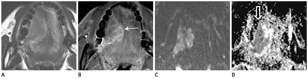
Fig. 1 A 47-year-old woman with SCC in the tongue. Axial T2- (A) and contrast enhanced T1-weighted images (B) show a mass in the right lateral tongue (arrow). DWI with b value of 1000 (C) reveals high signal intensity of the mass. The mean ADC value within the lesion measured 0.914 × 10⁻³ mm2/s on the ADC map image (D, open arrow). Note.-ADC = apparent diffusion coefficient, DWI = diffusion-weighted imaging, SCC = squamous cell carcinoma

Fig. 2 A 5-year-old male with lymphangioma in the left cheek. Axial T2- (A), T1-weighted images (B) reveal a mass with heterogenous signal intensity (arrows). The mass is enhanced in the central portion on contrast enhanced T1-weighted image (C). The mean ADC value of the mass measured 1.936 × 10⁻³ mm2/s on the ADC map image (D, open arrow). Note.-ADC = apparent diffusion coefficient
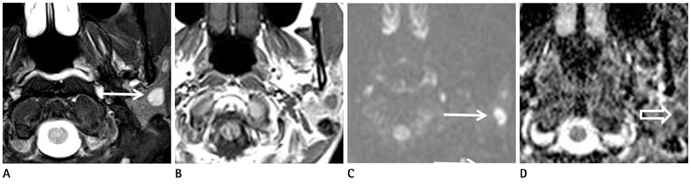
Fig. 3 A 47-year-old female with Warthin's tumor in the left parotid gland. Axial T2-weighted image (A) shows a round bright signal intensity mass (arrow). On T1-weighted image (B) the mass reveals low signal intensity. The mass represents high signal intensity on the DWI (C, arrow) and the ADC map image (D, open arrow). The ADC value of the lesion measured 0.731 × 10⁻³ mm2/s. Note.-ADC = apparent diffusion coefficient, DWI = diffusion-weighted imaging

Fig. 4 MR images of diffuse large B-cell lymphoma (A-C) in a 71-year-old woman and SCC (D-F) in a 58-year-old woman. In both patients, masses are located in the left nasopharynx (arrow), revealing hypointensity on T2-weighted image (A, D) and hyperintensity on DWI (B, E). The ADC value measured 0.526 × 10⁻³ mm2/s in lymphoma (C, open arrow), which was lower than that of patient with SCC (F, 0.877 × 10⁻³ mm2/s, open arrow). Note.-ADC = apparent diffusion coefficient, DWI = diffusion-weighted imaging, SCC = squamous cell carcinoma
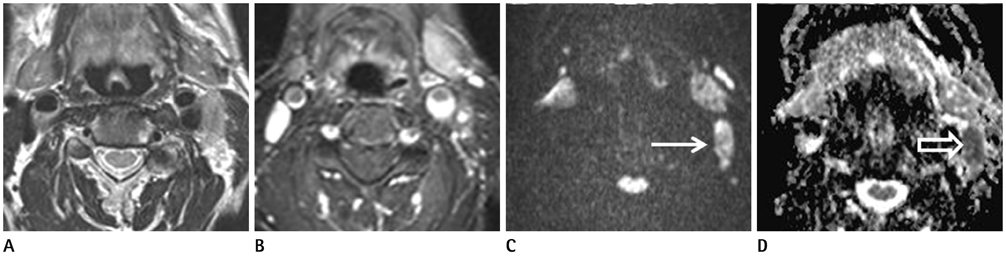
Fig. 5 A 64-year-old man with lymph nodal metastasis. Axial T2-weighted (A) and contrast enhanced T1-weighted images (B) reveal a 1-cm sized lymph node at the left level II, which is difficult to differentiate benign and malignant node. However, the node shows hyperintensity on DWI (C, arrow) and decreased ADC value (0.654 × 10⁻³ mm2/s) on the ADC map image (D, open arrow). Note.-ADC = apparent diffusion coefficient, DWI = diffusion-weighted imaging
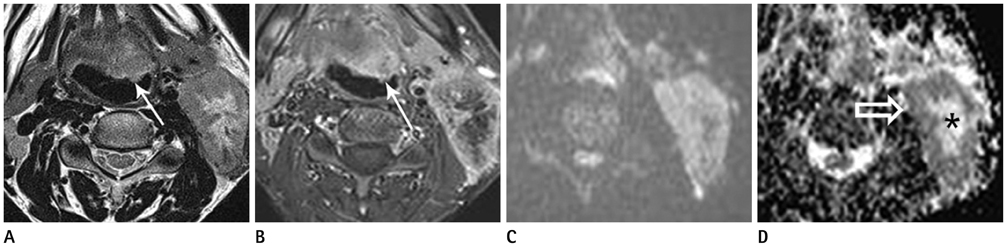
Fig. 6 A 49-year-old man with squamous cell cancer in the left tongue. Axial T2-weighted (A) and contrast enhanced T1-weighted images (B) show a malignant mass in the left tongue base (arrow), biopsy-proven SCC. A large metastatic lymphadenopathy is also seen in the left level II. The node shows hyperintensity with central hypointensity on DWI (C). The ADC map (D) reveals low signal intensity of the enhanced peripheral part of the tumor with low ADC value (0.987 × 10⁻³ mm2/s, open arrow) representing a viable portion of the tumor. The non-enhancing central part of the tumor shows high signal intensity with high ADC value (1.597 × 10⁻³ mm2/s, asterisk) representing a necrotic portion of the tumor. Note.-ADC = apparent diffusion coefficient, SCC = squamous cell carcinoma
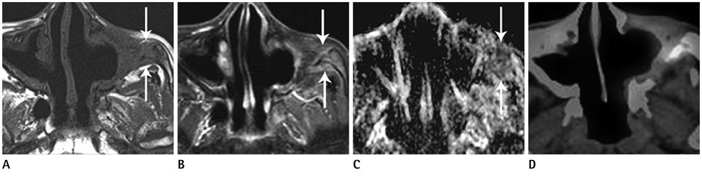
Fig. 7 A 71-year-old man with recurrent squamous cell cancer in the left maxillary sinus. Axial T1-weighted image (A) shows a mass at the lateral margin of previous resection site in the left maxillary sinus (arrows). On axial contrast T1-weighted image (B), the lesion is well-enhanced. The ADC map image (C) shows low signal intensity with a mean value of 0.581 × 10⁻³mm2/s, representing recurrence. Increased FDG uptake is noted in the lesion on PET/CT scan (D). Note.-ADC = apparent diffusion coefficient, FDG = fluorodeoxyglucose, PET/CT = positron emission tomography/CT

Fig. 8 A 54-year-old man with adenoid cystic carcinoma in the left hard palate. Initial contrast enhanced T1-weighted image (A) reveals an enhancing solid mass in the left palatal bone (arrows). Follow-up T1-weighted image (B) after post-radiation 1 1/2 year (B) shows markedly interval improving state. However, on the ADC map image at the same day (C), a focal area reveals diffusion restriction with a mean value of 0.74 × 10⁻³ mm2/s (open arrow), representing a remnant mass. The lesion is aggravated diffusely on the 5-month follow-up contrast enhanced T1-weighted image (D, arrows). Note.-ADC = apparent diffusion coefficient
Reference
-
1. Lell M, Baum U, Greess H, Nömayr A, Nkenke E, Koester M, et al. Head and neck tumors: imaging recurrent tumor and post-therapeutic changes with CT and MRI. Eur J Radiol. 2000; 33:239–247.2. Neil JJ. Diffusion imaging concepts for clinicians. J Magn Reson Imaging. 2008; 27:1–7.3. Malayeri AA, El Khouli RH, Zaheer A, Jacobs MA, Corona-Villalobos CP, Kamel IR, et al. Principles and applications of diffusion-weighted imaging in cancer detection, staging, and treatment follow-up. Radiographics. 2011; 31:1773–1791.4. Wang J, Takashima S, Takayama F, Kawakami S, Saito A, Matsushita T, et al. Head and neck lesions: characterization with diffusion-weighted echo-planar MR imaging. Radiology. 2001; 220:621–630.5. Sumi M, Sakihama N, Sumi T, Morikawa M, Uetani M, Kabasawa H, et al. Discrimination of metastatic cervical lymph nodes with diffusion-weighted MR imaging in patients with head and neck cancer. AJNR Am J Neuroradiol. 2003; 24:1627–1634.6. Srinivasan A, Dvorak R, Perni K, Rohrer S, Mukherji SK. Differentiation of benign and malignant pathology in the head and neck using 3T apparent diffusion coefficient values: early experience. AJNR Am J Neuroradiol. 2008; 29:40–44.7. Maeda M, Kato H, Sakuma H, Maier SE, Takeda K. Usefulness of the apparent diffusion coefficient in line scan diffusion-weighted imaging for distinguishing between squamous cell carcinomas and malignant lymphomas of the head and neck. AJNR Am J Neuroradiol. 2005; 26:1186–1192.8. Schaefer PW, Grant PE, Gonzalez RG. Diffusion-weighted MR imaging of the brain. Radiology. 2000; 217:331–345.9. Lang P, Johnston JO, Arenal-Romero F, Gooding CA. Advances in MR imaging of pediatric musculoskeletal neoplasms. Magn Reson Imaging Clin N Am. 1998; 6:579–604.10. Szafer A, Zhong J, Gore JC. Theoretical model for water diffusion in tissues. Magn Reson Med. 1995; 33:697–712.11. Le Bihan D, Breton E, Lallemand D, Aubin ML, Vignaud J, Laval-Jeantet M. Separation of diffusion and perfusion in intravoxel incoherent motion MR imaging. Radiology. 1988; 168:497–505.12. Kwon H, Kim HJ, Sol YL, Lee TH, Yeom JA, Kim AR. Usefulness of apparent diffusion coefficient values in the nasopharynx and the oropharynx: differentiation of benign and malignant lesions. J Korean Soc Radiol. 2012; 66:117–122.13. Sasaki M, Eida S, Sumi M, Nakamura T. Apparent diffusion coefficient mapping for sinonasal diseases: differentiation of benign and malignant lesions. AJNR Am J Neuroradiol. 2011; 32:1100–1106.14. Friedrich KM, Matzek W, Gentzsch S, Sulzbacher I, Czerny C, Herneth AM. Diffusion-weighted magnetic resonance imaging of head and neck squamous cell carcinomas. Eur J Radiol. 2008; 68:493–498.15. Habermann CR, Arndt C, Graessner J, Diestel L, Petersen KU, Reitmeier F, et al. Diffusion-weighted echo-planar MR imaging of primary parotid gland tumors: is a prediction of different histologic subtypes possible? AJNR Am J Neuroradiol. 2009; 30:591–596.16. Som PM, Brandwein MS. Salivary glands: anatomy and pathology. In : Som PM, Curtin HD, editors. Head and neck imaging. 4th ed. St. Louis, MO: Mosby;2003. p. 2005–2133.17. Sumi M, Ichikawa Y, Nakamura T. Diagnostic ability of apparent diffusion coefficients for lymphomas and carcinomas in the pharynx. Eur Radiol. 2007; 17:2631–2637.18. Curtin HD, Ishwaran H, Mancuso AA, Dalley RW, Caudry DJ, McNeil BJ. Comparison of CT and MR imaging in staging of neck metastases. Radiology. 1998; 207:123–130.19. van den Brekel MW. Lymph node metastases: CT and MRI. Eur J Radiol. 2000; 33:230–238.20. Abdel Razek AA, Soliman NY, Elkhamary S, Alsharaway MK, Tawfik A. Role of diffusion-weighted MR imaging in cervical lymphadenopathy. Eur Radiol. 2006; 16:1468–1477.21. Vandecaveye V, De Keyzer F, Vander Poorten V, Dirix P, Verbeken E, Nuyts S, et al. Head and neck squamous cell carcinoma: value of diffusion-weighted MR imaging for nodal staging. Radiology. 2009; 251:134–146.22. de Bondt RB, Hoeberigs MC, Nelemans PJ, Deserno WM, Peutz-Kootstra C, Kremer B, et al. Diagnostic accuracy and additional value of diffusion-weighted imaging for discrimination of malignant cervical lymph nodes in head and neck squamous cell carcinoma. Neuroradiology. 2009; 51:183–192.23. King AD, Ahuja AT, Yeung DK, Fong DK, Lee YY, Lei KI, et al. Malignant cervical lymphadenopathy: diagnostic accuracy of diffusion-weighted MR imaging. Radiology. 2007; 245:806–813.24. Koç O, Paksoy Y, Erayman I, Kivrak AS, Arbag H. Role of diffusion weighted MR in the discrimination diagnosis of the cystic and/or necrotic head and neck lesions. Eur J Radiol. 2007; 62:205–213.25. Razek AA, Megahed AS, Denewer A, Motamed A, Tawfik A, Nada N. Role of diffusion-weighted magnetic resonance imaging in differentiation between the viable and necrotic parts of head and neck tumors. Acta Radiol. 2008; 49:364–370.26. Tartaglino LM, Rao VM, Markiewicz DA. Imaging of radiation changes in the head and neck. Semin Roentgenol. 1994; 29:81–91.27. Terhaard CH, Bongers V, van Rijk PP, Hordijk GJ. F-18-fluoro-deoxy-glucose positron-emission tomography scanning in detection of local recurrence after radiotherapy for laryngeal/pharyngeal cancer. Head Neck. 2001; 23:933–941.28. Chisin R, Macapinlac HA. The indications of FDG-PET in neck oncology. Radiol Clin North Am. 2000; 38:999–1012.29. Hermans R, Pameijer FA, Mancuso AA, Parsons JT, Mendenhall WM. Laryngeal or hypopharyngeal squamous cell carcinoma: can follow-up CT after definitive radiation therapy be used to detect local failure earlier than clinical examination alone? Radiology. 2000; 214:683–687.30. Lai PH, Yang CF, Pan HB, Wu MT, Chu ST, Ger LP, et al. Recurrent inverted papilloma: diagnosis with pharmacokinetic dynamic gadolinium-enhanced MR imaging. AJNR Am J Neuroradiol. 1999; 20:1445–1451.31. Abdel Razek AA, Kandeel AY, Soliman N, El-shenshawy HM, Kamel Y, Nada N, et al. Role of diffusion-weighted echo-planar MR imaging in differentiation of residual or recurrent head and neck tumors and posttreatment changes. AJNR Am J Neuroradiol. 2007; 28:1146–1152.32. Baur A, Huber A, Arbogast S, Dürr HR, Zysk S, Wendtner C, et al. Diffusion-weighted imaging of tumor recurrencies and posttherapeutical soft-tissue changes in humans. Eur Radiol. 2001; 11:828–833.33. Maier CF, Paran Y, Bendel P, Rutt BK, Degani H. Quantitative diffusion imaging in implanted human breast tumors. Magn Reson Med. 1997; 37:576–581.34. Eis M, Els T, Hoehn-Berlage M. High resolution quantitative relaxation and diffusion MRI of three different experimental brain tumors in rat. Magn Reson Med. 1995; 34:835–844.35. Chenevert TL, McKeever PE, Ross BD. Monitoring early response of experimental brain tumors to therapy using diffusion magnetic resonance imaging. Clin Cancer Res. 1997; 3:1457–1466.36. Poptani H, Puumalainen AM, Gröhn OH, Loimas S, Kainulainen R, Ylä-Herttuala S, et al. Monitoring thymidine kinase and ganciclovir-induced changes in rat malignant glioma in vivo by nuclear magnetic resonance imaging. Cancer Gene Ther. 1998; 5:101–109.37. Tsuruda JS, Chew WM, Moseley ME, Norman D. Diffusion-weighted MR imaging of the brain: value of differentiating between extraaxial cysts and epidermoid tumors. AJR Am J Roentgenol. 1990; 155:1059–1065. discussion 1066-1068.38. Brunberg JA, Chenevert TL, McKeever PE, Ross DA, Junck LR, Muraszko KM, et al. In vivo MR determination of water diffusion coefficients and diffusion anisotropy: correlation with structural alteration in gliomas of the cerebral hemispheres. AJNR Am J Neuroradiol. 1995; 16:361–371.39. Chenevert TL, Stegman LD, Taylor JM, Robertson PL, Greenberg HS, Rehemtulla A, et al. Diffusion magnetic resonance imaging: an early surrogate marker of therapeutic efficacy in brain tumors. J Natl Cancer Inst. 2000; 92:2029–2036.40. Mardor Y, Pfeffer R, Spiegelmann R, Roth Y, Maier SE, Nissim O, et al. Early detection of response to radiation therapy in patients with brain malignancies using conventional and high b-value diffusion-weighted magnetic resonance imaging. J Clin Oncol. 2003; 21:1094–1100.41. Zhao M, Pipe JG, Bonnett J, Evelhoch JL. Early detection of treatment response by diffusion-weighted 1H-NMR spectroscopy in a murine tumour in vivo. Br J Cancer. 1996; 73:61–64.42. Stegman LD, Rehemtulla A, Hamstra DA, Rice DJ, Jonas SJ, Stout KL, et al. Diffusion MRI detects early events in the response of a glioma model to the yeast cytosine deaminase gene therapy strategy. Gene Ther. 2000; 7:1005–1010.43. Moffat BA, Chenevert TL, Lawrence TS, Meyer CR, Johnson TD, Dong Q, et al. Functional diffusion map: a noninvasive MRI biomarker for early stratification of clinical brain tumor response. Proc Natl Acad Sci U S A. 2005; 102:5524–5529.44. Lee KC, Moffat BA, Schott AF, Layman R, Ellingworth S, Juliar R, et al. Prospective early response imaging biomarker for neoadjuvant breast cancer chemotherapy. Clin Cancer Res. 2007; 13(2 Pt 1):443–450.45. McVeigh PZ, Syed AM, Milosevic M, Fyles A, Haider MA. Diffusion-weighted MRI in cervical cancer. Eur Radiol. 2008; 18:1058–1064.46. Kim S, Loevner L, Quon H, Sherman E, Weinstein G, Kilger A, et al. Diffusion-weighted magnetic resonance imaging for predicting and detecting early response to chemoradiation therapy of squamous cell carcinomas of the head and neck. Clin Cancer Res. 2009; 15:986–994.47. Chinnaiyan AM, Prasad U, Shankar S, Hamstra DA, Shanaiah M, Chenevert TL, et al. Combined effect of tumor necrosis factor-related apoptosis-inducing ligand and ionizing radiation in breast cancer therapy. Proc Natl Acad Sci U S A. 2000; 97:1754–1759.48. Kauppinen RA. Monitoring cytotoxic tumour treatment response by diffusion magnetic resonance imaging and proton spectroscopy. NMR Biomed. 2002; 15:6–17.49. Cao Y, Popovtzer A, Li D, Chepeha DB, Moyer JS, Prince ME, et al. Early prediction of outcome in advanced head-and-neck cancer based on tumor blood volume alterations during therapy: a prospective study. Int J Radiat Oncol Biol Phys. 2008; 72:1287–1290.50. Schmitt P, Kotas M, Tobermann A, Haase A, Flentje M. Quantitative tissue perfusion measurements in head and neck carcinoma patients before and during radiation therapy with a non-invasive MR imaging spin-labeling technique. Radiother Oncol. 2003; 67:27–34.51. Robinson SP, Collingridge DR, Howe FA, Rodrigues LM, Chaplin DJ, Griffiths JR. Tumour response to hypercapnia and hyperoxia monitored by FLOOD magnetic resonance imaging. NMR Biomed. 1999; 12:98–106.52. Zima A, Carlos R, Gandhi D, Case I, Teknos T, Mukherji SK. Can pretreatment CT perfusion predict response of advanced squamous cell carcinoma of the upper aerodigestive tract treated with induction chemotherapy? AJNR Am J Neuroradiol. 2007; 28:328–334.53. Pruessmann KP, Weiger M, Scheidegger MB, Boesiger P. SENSE: sensitivity encoding for fast MRI. Magn Reson Med. 1999; 42:952–962.54. Willinek WA, Gieseke J, von Falkenhausen M, Neuen B, Schild HH, Kuhl CK. Sensitivity encoding for fast MR imaging of the brain in patients with stroke. Radiology. 2003; 228:669–675.55. Yoshino N, Yamada I, Ohbayashi N, Honda E, Ida M, Kurabayashi T, et al. Salivary glands and lesions: evaluation of apparent diffusion coefficients with split-echo diffusion-weighted MR imaging--initial results. Radiology. 2001; 221:837–842.56. Kito S, Morimoto Y, Tanaka T, Tominaga K, Habu M, Kurokawa H, et al. Utility of diffusion-weighted images using fast asymmetric spin-echo sequences for detection of abscess formation in the head and neck region. Oral Surg Oral Med Oral Pathol Oral Radiol Endod. 2006; 101:231–238.57. Grieve SM, Blamire AM, Styles P. Elimination of Nyquist ghosting caused by read-out to phase-encode gradient cross-terms in EPI. Magn Reson Med. 2002; 47:337–343.58. Vandecaveye V, De Keyzer F, Nuyts S, Deraedt K, Dirix P, Hamaekers P, et al. Detection of head and neck squamous cell carcinoma with diffusion weighted MRI after (chemo)radiotherapy: correlation between radiologic and histopathologic findings. Int J Radiat Oncol Biol Phys. 2007; 67:960–971.
- Full Text Links
-
- Actions
-
Cited
- CITED
-
- Close
- Share
-
- Similar articles
-
- Advanced Magnetic Resonance Imaging for Pediatric Brain Tumors: Current Imaging Techniques and Interpretation Algorithms
- The Value of PROPELLER Diffusion-Weighted Image in the Detection of Cholesteatoma
- Current Applications and Future Perspectives of Brain Tumor Imaging
- Diffusion-Weighted MR Imaging of Unicystic Odontogenic Tumors for Differentiation of Unicystic Ameloblastomas from Keratocystic Odontogenic Tumors
- Diffusion-weighted Magnetic Resonance Imaging for Predicting Response to Chemoradiation Therapy for Head and Neck Squamous Cell Carcinoma: A Systematic Review
