Korean J Radiol.
2019 Apr;20(4):649-661. 10.3348/kjr.2018.0446.
Diffusion-weighted Magnetic Resonance Imaging for Predicting Response to Chemoradiation Therapy for Head and Neck Squamous Cell Carcinoma: A Systematic Review
- Affiliations
-
- 1Department of Radiology and Research Institute of Radiology, University of Ulsan College of Medicine, Asan Medical Center, Seoul, Korea. jehee23@gmail.com
- 2Department of Radiology, Namwon Medical Center, Namwon, Korea.
- KMID: 2440488
- DOI: http://doi.org/10.3348/kjr.2018.0446
Abstract
OBJECTIVE
To systematically review the evaluation of the diagnostic accuracy of pre-treatment apparent diffusion coefficient (ADC) and change in ADC during the intra- or post-treatment period, for the prediction of locoregional failure in patients with head and neck squamous cell carcinoma (HNSCC).
MATERIALS AND METHODS
Ovid-MEDLINE and Embase databases were searched up to September 8, 2018, for studies on the use of diffusion-weighted magnetic resonance imaging for the prediction of locoregional treatment response in patients with HNSCC treated with chemoradiation or radiation therapy. Risk of bias was assessed by using the Quality Assessment Tool for Diagnostic Accuracy Studies-2.
RESULTS
Twelve studies were included in the systematic review, and diagnostic accuracy assessment was performed using seven studies. High pre-treatment ADC showed inconsistent results with the tendency for locoregional failure, whereas all studies evaluating changes in ADC showed consistent results of a lower rise in ADC in patients with locoregional failure compared to those with locoregional control. The sensitivities and specificities of pre-treatment ADC and change in ADC for predicting locoregional failure were relatively high (range: 50-100% and 79-96%, 75-100% and 69-95%, respectively). Meta-analytic pooling was not performed due to the apparent heterogeneity in these values.
CONCLUSION
High pre-treatment ADC and low rise in early intra-treatment or post-treatment ADC with chemoradiation, could be indicators of locoregional failure in patients with HNSCC. However, as the studies are few, heterogeneous, and at high risk for bias, the sensitivity and specificity of these parameters for predicting the treatment response are yet to be determined.
Keyword
MeSH Terms
Figure
-
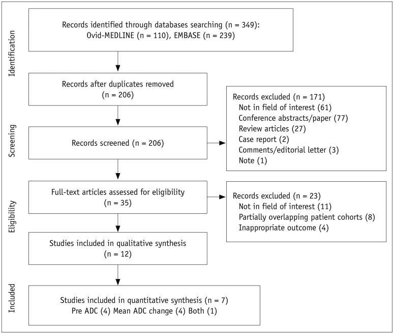
Fig. 1 Flow diagram of study selection process.ADC = apparent diffusion coefficient
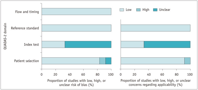
Fig. 2 QUADAS-2 criteria for included studies.QUADAS-2 = Quality Assessment of Diagnostic Accuracy Studies-2
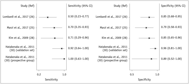
Fig. 3 Forest plots of sensitivity and specificity of pre-treatment ADC for prediction of locoregional recurrence.Horizontal lines indicate 95% CIs of individual studies. CI = confidence interval, Ref = reference
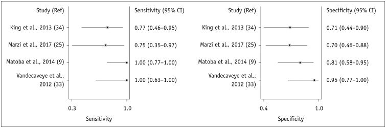
Fig. 4 Forest plots of sensitivity and specificity of change in ADC for prediction of locoregional recurrence.Horizontal lines indicate 95% CIs of individual studies.
Reference
-
1. Bonner JA, Harari PM, Giralt J, Azarnia N, Shin DM, Cohen RB, et al. Radiotherapy plus cetuximab for squamous-cell carcinoma of the head and neck. N Engl J Med. 2006; 354:567–578. PMID: 16467544.
Article2. Argiris A, Karamouzis MV, Raben D, Ferris RL. Head and neck cancer. Lancet. 2008; 371:1695–1709. PMID: 18486742.
Article3. Chiesa F, Mauri S, Tradati N, Calabrese L, Giugliano G, Ansarin M, et al. Surfing prognostic factors in head and neck cancer at the millennium. Oral Oncol. 1999; 35:590–596. PMID: 10705095.
Article4. Ang KK, Trotti A, Brown BW, Garden AS, Foote RL, Morrison WH, et al. Randomized trial addressing risk features and time factors of surgery plus radiotherapy in advanced head-and-neck cancer. Int J Radiat Oncol Biol Phys. 2001; 51:571–578. PMID: 11597795.
Article5. Agra IM, Carvalho AL, Ulbrich FS, de Campos OD, Martins EP, Magrin J, et al. Prognostic factors in salvage surgery for recurrent oral and oropharyngeal cancer. Head Neck. 2006; 28:107–113. PMID: 16388526.
Article6. Carvalho AL, Magrin J, Kowalski LP. Sites of recurrence in oral and oropharyngeal cancers according to the treatment approach. Oral Dis. 2003; 9:112–118. PMID: 12945592.
Article7. Goodwin WJ Jr. Salvage surgery for patients with recurrent squamous cell carcinoma of the upper aerodigestive tract: when do the ends justify the means? Laryngoscope. 2000; 110:1–18.
Article8. Kowalski LP, Bagietto R, Lara JR, Santos RL, Silva JF Jr, Magrin J. Prognostic significance of the distribution of neck node metastasis from oral carcinoma. Head Neck. 2000; 22:207–214. PMID: 10748442.
Article9. Matoba M, Tuji H, Shimode Y, Toyoda I, Kuginuki Y, Miwa K, et al. Fractional change in apparent diffusion coefficient as an imaging biomarker for predicting treatment response in head and neck cancer treated with chemoradiotherapy. AJNR Am J Neuroradiol. 2014; 35:379–385. PMID: 24029391.
Article10. Newman JP, Terris DJ, Pinto HA, Fee WE Jr, Goode RL, Goffinet DR. Surgical morbidity of neck dissection after chemoradiotherapy in advanced head and neck cancer. Ann Otol Rhinol Laryngol. 1997; 106:117–122. PMID: 9041815.
Article11. Lavertu P, Bonafede JP, Adelstein DJ, Saxton JP, Strome M, Wanamaker JR, et al. Comparison of surgical complications after organ-preservation therapy in patients with stage III or IV squamous cell head and neck cancer. Arch Otolaryngol Head Neck Surg. 1998; 124:401–406. PMID: 9559686.
Article12. Davuluri R, Krase JM, Cui H, Goyal UD, Cheung MK, Hsu CC, et al. Image guided volumetric response during chemoradiotherapy for head and neck squamous cell carcinoma as a predictor of disease outcomes. Am J Otolaryngol. 2016; 37:304–310. PMID: 27105977.13. Tang C, Fuller CD, Garden AS, Awan MJ, Colen RR, Morrison WH, et al. Characteristics and kinetics of cervical lymph node regression after radiation therapy for human papillomavirus-associated oropharyngeal carcinoma: quantitative image analysis of post-radiotherapy response. Oral Oncol. 2015; 51:195–201. PMID: 25444304.
Article14. Belli ML, Fiorino C, Zerbetto F, Raso R, Broggi S, Chiara A, et al. Early volume variation of positive lymph nodes assessed by in-room mega voltage CT images predicts risk of loco-regional relapses in head and neck cancer patients treated with intensity-modulated radiotherapy. Acta Oncol. 2015; 54:1490–1495. PMID: 26203925.
Article15. Ojiri H, Mendenhall WM, Mancuso AA. CT findings at the primary site of oropharyngeal squamous cell carcinoma within 6-8 weeks after definitive radiotherapy as predictors of primary site control. Int J Radiat Oncol Biol Phys. 2002; 52:748–754. PMID: 11849798.
Article16. Hermans R, Pameijer FA, Mancuso AA, Parsons JT, Mendenhall WM. Laryngeal or hypopharyngeal squamous cell carcinoma: can follow-up CT after definitive radiation therapy be used to detect local failure earlier than clinical examination alone? Radiology. 2000; 214:683–687. PMID: 10715030.
Article17. King AD, Keung CK, Yu KH, Mo FK, Bhatia KS, Yeung DK, et al. T2-weighted MR imaging early after chemoradiotherapy to evaluate treatment response in head and neck squamous cell carcinoma. AJNR Am J Neuroradiol. 2013; 34:1237–1241. PMID: 23306012.
Article18. Lell M, Baum U, Greess H, Nomayr A, Nkenke E, Koester M, et al. Head and neck tumors: imaging recurrent tumor and post-therapeutic changes with CT and MRI. Eur J Radiol. 2000; 33:239–247. PMID: 10699740.
Article19. Sanguineti G, Ricchetti F, Wu B, Agrawal N, Gourin C, Agbahiwe H, et al. Volumetric change of human papillomavirus-related neck lymph nodes before, during, and shortly after intensity-modulated radiation therapy. Head Neck. 2012; 34:1640–1647. PMID: 22267196.
Article20. Munck JN, Cvitkovic E, Piekarski JD, Benhamou E, Recondo G, Bachouchi M, et al. Computed tomographic density of metastatic lymph nodes as a treatment-related prognostic factor in advanced head and neck cancer. J Natl Cancer Inst. 1991; 83:569–575. PMID: 1848639.
Article21. McCollum AD, Burrell SC, Haddad RI, Norris CM, Tishler RB, Case MA, et al. Positron emission tomography with 18F-fluorodeoxyglucose to predict pathologic response after induction chemotherapy and definitive chemoradiotherapy in head and neck cancer. Head Neck. 2004; 26:890–896. PMID: 15390197.22. King AD, Mo FK, Yu KH, Yeung DK, Zhou H, Bhatia KS, et al. Squamous cell carcinoma of the head and neck: diffusion-weighted MR imaging for prediction and monitoring of treatment response. Eur Radiol. 2010; 20:2213–2220. PMID: 20309553.
Article23. Srinivasan A, Chenevert TL, Dwamena BA, Eisbruch A, Watcharotone K, Myles JD, et al. Utility of pretreatment mean apparent diffusion coefficient and apparent diffusion coefficient histograms in prediction of outcome to chemoradiation in head and neck squamous cell carcinoma. J Comput Assist Tomogr. 2012; 36:131–137. PMID: 22261783.
Article24. Ng SH, Lin CY, Chan SC, Lin YC, Yen TC, Liao CT, et al. Clinical utility of multimodality imaging with dynamic contrast-enhanced MRI, diffusion-weighted MRI, and 18F-FDG PET/CT for the prediction of neck control in oropharyngeal or hypopharyngeal squamous cell carcinoma treated with chemoradiation. PLoS One. 2014; 9:e115933. PMID: 25531391.
Article25. Marzi S, Piludu F, Sanguineti G, Marucci L, Farneti A, Terrenato I, et al. The prediction of the treatment response of cervical nodes using intravoxel incoherent motion diffusion-weighted imaging. Eur J Radiol. 2017; 92:93–102. PMID: 28624026.
Article26. Lombardi M, Cascone T, Guenzi E, Stecco A, Buemi F, Krengli M, et al. Predictive value of pre-treatment apparent diffusion coefficient (ADC) in radio-chemiotherapy treated head and neck squamous cell carcinoma. Radiol Med. 2017; 122:345–352. PMID: 28188603.
Article27. Lambrecht M, Van Calster B, Vandecaveye V, De Keyzer F, Roebben I, Hermans R, et al. Integrating pretreatment diffusion weighted MRI into a multivariable prognostic model for head and neck squamous cell carcinoma. Radiother Oncol. 2014; 110:429–434. PMID: 24630535.
Article28. Kim S, Loevner L, Quon H, Sherman E, Weinstein G, Kilger A, et al. Diffusion-weighted magnetic resonance imaging for predicting and detecting early response to chemoradiation therapy of squamous cell carcinomas of the head and neck. Clin Cancer Res. 2009; 15:986–994. PMID: 19188170.
Article29. Hauser T, Essig M, Jensen A, Gerigk L, Laun FB, Munter M, et al. Characterization and therapy monitoring of head and neck carcinomas using diffusion-imaging-based intravoxel incoherent motion parameters-preliminary results. Neuroradiology. 2013; 55:527–536. PMID: 23417120.
Article30. Hatakenaka M, Shioyama Y, Nakamura K, Yabuuchi H, Matsuo Y, Sunami S, et al. Apparent diffusion coefficient calculated with relatively high b-values correlates with local failure of head and neck squamous cell carcinoma treated with radiotherapy. AJNR Am J Neuroradiol. 2011; 32:1904–1910. PMID: 21778248.
Article31. Hatakenaka M, Nakamura K, Yabuuchi H, Shioyama Y, Matsuo Y, Ohnishi K, et al. Pretreatment apparent diffusion coefficient of the primary lesion correlates with local failure in head-and-neck cancer treated with chemoradiotherapy or radiotherapy. Int J Radiat Oncol Biol Phys. 2011; 81:339–345. PMID: 20832179.
Article32. Hatakenaka M, Nakamura K, Yabuuchi H, Shioyama Y, Matsuo Y, Kamitani T, et al. Apparent diffusion coefficient is a prognostic factor of head and neck squamous cell carcinoma treated with radiotherapy. Jpn J Radiol. 2014; 32:80–89. PMID: 24408077.
Article33. Vandecaveye V, Dirix P, De Keyzer F, Op de Beeck K, Vander Poorten V, Hauben E, et al. Diffusion-weighted magnetic resonance imaging early after chemoradiotherapy to monitor treatment response in head-and-neck squamous cell carcinoma. Int J Radiat Oncol Biol Phys. 2012; 82:1098–1107. PMID: 21514067.
Article34. King AD, Chow KK, Yu KH, Mo FK, Yeung DK, Yuan J, et al. Head and neck squamous cell carcinoma: diagnostic performance of diffusion-weighted MR imaging for the prediction of treatment response. Radiology. 2013; 266:531–538. PMID: 23151830.
Article35. Wong KH, Panek R, Welsh L, McQuaid D, Dunlop A, Riddell A, et al. The predictive value of early assessment after 1 cycle of induction chemotherapy with 18F-FDG PET/CT and diffusion-weighted mri for response to radical chemoradiotherapy in head and neck squamous cell carcinoma. J Nucl Med. 2016; 57:1843–1850. PMID: 27417648.
Article36. Schouten CS, de Bree R, van der Putten L, Noij DP, Hoekstra OS, Comans EF, et al. Diffusion-weighted EPI- and HASTE-MRI and 18F-FDG-PET-CT early during chemoradiotherapy in advanced head and neck cancer. Quant Imaging Med Surg. 2014; 4:239–250. PMID: 25202659.37. Paudyal R, Oh JH, Riaz N, Venigalla P, Li J, Hatzoglou V, et al. Intravoxel incoherent motion diffusion-weighted MRI during chemoradiation therapy to characterize and monitor treatment response in human papillomavirus head and neck squamous cell carcinoma. J Magn Reson Imaging. 2017; 45:1013–1023. PMID: 27862553.
Article38. Galbán CJ, Mukherji SK, Chenevert TL, Meyer CR, Hamstra DA, Bland PH, et al. A feasibility study of parametric response map analysis of diffusion-weighted magnetic resonance imaging scans of head and neck cancer patients for providing early detection of therapeutic efficacy. Transl Oncol. 2009; 2:184–190. PMID: 19701503.
Article39. Aramburu Núñez D, Lopez MA, Mera IM, Salvador GF, Dave A, Hatzoglou V, et al. Multimodality functional imaging using DW-MRI and 18F-FDG-PET/CT during radiation therapy for human papillomavirus negative head and neck squamous cell carcinoma: Meixoeiro Hospital of Vigo Experience. World J Radiol. 2017; 9:17–26. PMID: 28144403.40. Hauser T, Essig M, Jensen A, Laun FB, Munter M, Maier-Hein KH, et al. Prediction of treatment response in head and neck carcinomas using IVIM-DWI: evaluation of lymph node metastasis. Eur J Radiol. 2014; 83:783–787. PMID: 24631600.
Article41. Whiting PF, Rutjes AW, Westwood ME, Mallett S, Deeks JJ, Reitsma JB, et al. QUADAS-2: a revised tool for the quality assessment of diagnostic accuracy studies. Ann Intern Med. 2011; 155:529–536. PMID: 22007046.
Article42. Deville WL, Buntinx F, Bouter LM, Montori VM, de Vet HC, van der Windt DA, et al. Conducting systematic reviews of diagnostic studies: didactic guidelines. BMC Med Res Methodol. 2002; 2:9. PMID: 12097142.
Article43. Vandecaveye V, Dirix P, De Keyzer F, de Beeck KO, Vander Poorten V, Roebben I, et al. Predictive value of diffusion-weighted magnetic resonance imaging during chemoradiotherapy for head and neck squamous cell carcinoma. Eur Radiol. 2010; 20:1703–1714. PMID: 20179939.
Article44. King AD, Thoeny HC. Functional MRI for the prediction of treatment response in head and neck squamous cell carcinoma: potential and limitations. Cancer Imaging. 2016; 16:23. PMID: 27542718.
Article45. Driessen JP, Caldas-Magalhaes J, Janssen LM, Pameijer FA, Kooij N, Terhaard CH, et al. Diffusion-weighted MR imaging in laryngeal and hypopharyngeal carcinoma: association between apparent diffusion coefficient and histologic findings. Radiology. 2014; 272:456–463. PMID: 24749712.
Article46. Driessen JP, van Bemmel AJ, van Kempen PM, Janssen LM, Terhaard CH, Pameijer FA, et al. Correlation of human papillomavirus status with apparent diffusion coefficient of diffusion-weighted MRI in head and neck squamous cell carcinomas. Head Neck. 2016; 38(Suppl 1):E613–E618. PMID: 25783872.
Article47. Nakahira M, Saito N, Yamaguchi H, Kuba K, Sugasawa M. Use of quantitative diffusion-weighted magnetic resonance imaging to predict human papilloma virus status in patients with oropharyngeal squamous cell carcinoma. Eur Arch Otorhinolaryngol. 2014; 271:1219–1225. PMID: 23880924.
Article48. Abdel Razek AA, Kandeel AY, Soliman N, El-shenshawy HM, Kamel Y, Nada N, et al. Role of diffusion-weighted echo-planar MR imaging in differentiation of residual or recurrent head and neck tumors and posttreatment changes. AJNR Am J Neuroradiol. 2007; 28:1146–1152. PMID: 17569975.49. Pickles MD, Gibbs P, Lowry M, Turnbull LW. Diffusion changes precede size reduction in neoadjuvant treatment of breast cancer. Magn Reson Imaging. 2006; 24:843–847. PMID: 16916701.
Article50. Uhl M, Saueressig U, van Buiren M, Kontny U, Niemeyer C, Kohler G, et al. Osteosarcoma: preliminary results of in vivo assessment of tumor necrosis after chemotherapy with diffusion- and perfusion-weighted magnetic resonance imaging. Invest Radiol. 2006; 41:618–623. PMID: 16829744.51. Mardor Y, Pfeffer R, Spiegelmann R, Roth Y, Maier SE, Nissim O, et al. Early detection of response to radiation therapy in patients with brain malignancies using conventional and high b-value diffusion-weighted magnetic resonance imaging. J Clin Oncol. 2003; 21:1094–1100. PMID: 12637476.
Article52. Chen YG, Chen MQ, Guo YY, Li SC, Wu JX, Xu BH. Apparent diffusion coefficient predicts pathology complete response of rectal cancer treated with neoadjuvant chemoradiotherapy. PLoS One. 2016; 11:e0153944. PMID: 27100991.
Article53. Onal C, Erbay G, Guler OC. Treatment response evaluation using the mean apparent diffusion coefficient in cervical cancer patients treated with definitive chemoradiotherapy. J Magn Reson Imaging. 2016; 44:1010–1019. PMID: 26919800.
Article54. Argiris A, Stenson KM, Brockstein BE, Mittal BB, Pelzer H, Kies MS, et al. Neck dissection in the combined-modality therapy of patients with locoregionally advanced head and neck cancer. Head Neck. 2004; 26:447–455. PMID: 15122662.
Article55. Brizel DM, Prosnitz RG, Hunter S, Fisher SR, Clough RL, Downey MA, et al. Necessity for adjuvant neck dissection in setting of concurrent chemoradiation for advanced head-and-neck cancer. Int J Radiat Oncol Biol Phys. 2004; 58:1418–1423. PMID: 15050318.
Article
- Full Text Links
-
- Actions
-
Cited
- CITED
-
- Close
- Share
-
- Similar articles
-
- Squamous Cell Carcinoma of the Pancreas: A Case Report
- Coexistence of Intracranial Squamous Cell Carcinoma and Epidermoid Cyst: a Case with Consecutive Imaging Findings
- Diffusion-Weighted Magnetic Resonance Imaging of Spine
- RE: Distinguishing between Renal Cell Carcinoma and Fat Poor Angiomyolipoma in Diffusion-Weighted Imaging
- The Value of PROPELLER Diffusion-Weighted Image in the Detection of Cholesteatoma
