Diffusion-Weighted MR Imaging of Unicystic Odontogenic Tumors for Differentiation of Unicystic Ameloblastomas from Keratocystic Odontogenic Tumors
- Affiliations
-
- 1Department of Interventional Radiology, Ninth People's Hospital, Shanghai Jiao Tong University, Shanghai 200011, China. fanxindong@aliyun.com
- 2Department of Oral & Maxillofacial Surgery, Ninth People's Hospital, Shanghai Jiao Tong University, Shanghai 200011, China.
- KMID: 2425112
- DOI: http://doi.org/10.3348/kjr.2018.19.1.79
Abstract
OBJECTIVE
Differentiating unicystic ameloblastomas from keratocystic odontogenic tumors (KCOT) is necessary for the planning of different treatment strategies; however, it is difficult based on conventional CT and MR sequences alone. The purpose of this study was to investigate the utility of diffusion-weighted imaging (DWI) and apparent diffusion coefficients (ADCs) in the differentiation of the two tumors.
MATERIALS AND METHODS
We prospectively studied 40 patients with odontogenic cysts and tumors of the maxillomandibular region using conventional MR imaging and DWI. ADCs were measured using 2 b factors (500 and 1000).
RESULTS
Unicystic ameloblastomas (n = 11) showed free diffusion on DWI and a mean ADC value of 2.309 ± 0.17 × 10⁻³ mm2/s. KCOT (n = 15) showed restricted diffusion on DWI with a mean ADC value of 0.923 ± 0.20 × 10⁻³ mm2/s. The ADC values of unicystic ameloblastomas were significantly higher than those of KCOT (p < 0.001, Mann-Whitney U-test). An ADC cut-off value of 2.0 × 10⁻³ mm2/s to differentiate KCOT and unicystic ameloblastomas resulted in a 100% sensitivity and 100% specificity. Dentigerous cysts (n = 3) showed restricted diffusion on DWI and similar ADC values (1.257 ± 0.05 × 10⁻³ mm2/s) to those of KCOT.
CONCLUSION
Diffusion-weighted imaging and ADC determination can be used as an adjuvant tool to differentiate between unicystic ameloblastomas and KCOT, although the ADC values of dentigerous cysts overlap with those of KCOT.
Keyword
MeSH Terms
Figure
-
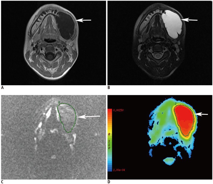
Fig. 1 12-year-old male patient with unicystic ameloblastoma at left mandibular region.A. Axial contrast-enhanced T1-weighted image shows unilocular cystic lesion (arrow) with rim enhancement. B. Axial fat-suppressed T2-weighted image shows lesion (arrow) with high signal intensity. C. Diffusion-weighted image at b = 1000 s/mm2 shows loss of signal within lesion (arrow).D. Axial color ADC map shows lesion (arrow) with high ADC (2.013 × 10⁻³ mm2/s). ADC = apparent diffusion coefficient

Fig. 2 25-year-old male patient with keratocystic odontogenic tumors at right mandibular region.A. Axial contrast-enhanced T1-weighted image shows unilocular cystic lesion (arrow) with rim enhancement in mandibular ramus. B. Axial fat-suppressed T2-weighted image shows lesion (arrow) with high signal intensity. C. Diffusion-weighted image at b = 1000 s/mm2 shows that lesion has retained signal (arrow). D. Axial color ADC map shows lesion (arrow) with low ADC (1.02 × 103 mm2/s).

Fig. 3 Box-and-whisker plot for comparing apparent diffusion coefficient values of unicystic ameloblastomas, KCOT and dentigerous cysts. p, Mann-Whitney U test.Bottom and top of each box and whisker plot are first and third quartiles, and band inside box is second quartile. Ends of whiskers represent minimum and maximum of all data. All data are included between whiskers. KCOT = keratocystic odontogenic tumors
Reference
-
1. Minami M, Kaneda T, Yamamoto H, Ozawa K, Itai Y, Ozawa M, et al. Ameloblastoma in the maxillomandibular region: MR imaging. Radiology. 1992; 184:389–393. PMID: 1620834.
Article2. Minami M, Kaneda T, Ozawa K, Yamamoto H, Itai Y, Ozawa M, et al. Cystic lesions of the maxillomandibular region: MR imaging distinction of odontogenic keratocysts and ameloblastomas from other cysts. AJR Am J Roentgenol. 1996; 166:943–949. PMID: 8610578.
Article3. Fujita M, Matsuzaki H, Yanagi Y, Hara M, Katase N, Hisatomi M, et al. Diagnostic value of MRI for odontogenic tumours. Dentomaxillofac Radiol. 2013; 42:20120265. PMID: 23468124.
Article4. Carlson ER, Marx RE. The ameloblastoma: primary, curative surgical management. J Oral Maxillofac Surg. 2006; 64:484–494. PMID: 16487813.
Article5. Ecker J, Horst RT, Koslovsky D. Current role of carnoy's solution in treating keratocystic odontogenic tumors. J Oral Maxillofac Surg. 2016; 74:278–282. PMID: 26272006.
Article6. Rosenstein T, Pogrel MA, Smith RA, Regezi JA. Cystic ameloblastoma--behavior and treatment of 21 cases. J Oral Maxillofac Surg. 2001; 59:1311–1316. discussion 1316-1318. PMID: 11688034.
Article7. Le Bihan D, Turner R, Douek P, Patronas N. Diffusion MR imaging: clinical applications. AJR Am J Roentgenol. 1992; 159:591–599. PMID: 1503032.
Article8. Lim HK, Lee JH, Baek HJ, Kim N, Lee H, Park JW, et al. Is diffusion-weighted MRI useful for differentiation of small non-necrotic cervical lymph nodes in patients with head and neck malignancies? Korean J Radiol. 2014; 15:810–816. PMID: 25469094.
Article9. Sumi M, Ichikawa Y, Katayama I, Tashiro S, Nakamura T. Diffusion-weighted MR imaging of ameloblastomas and keratocystic odontogenic tumors: differentiation by apparent diffusion coefficients of cystic lesions. AJNR Am J Neuroradiol. 2008; 29:1897–1901. PMID: 18719033.
Article10. Srinivasan K, Seith Bhalla A, Sharma R, Kumar A, Roychoudhury A, Bhutia O. Diffusion-weighted imaging in the evaluation of odontogenic cysts and tumours. Br J Radiol. 2012; 85:e864–e870. PMID: 22553294.
Article11. Kaneda T, Minami M, Kurabayashi T. Benign odontogenic tumors of the mandible and maxilla. Neuroimaging Clin N Am. 2003; 13:495–507. PMID: 14631687.
Article12. Ackermann GL, Altini M, Shear M. The unicystic ameloblastoma: a clinicopathological study of 57 cases. J Oral Pathol. 1988; 17:541–546. PMID: 3150441.
Article13. Probst FA, Probst M, Pautke Ch, Kaltsi E, Otto S, Schiel S, et al. Magnetic resonance imaging: a useful tool to distinguish between keratocystic odontogenic tumours and odontogenic cysts. Br J Oral Maxillofac Surg. 2015; 53:217–222. PMID: 25554593.
Article14. Konouchi H, Asaumi J, Yanagi Y, Hisatomi M, Kawai N, Matsuzaki H, et al. Usefulness of contrast enhanced-MRI in the diagnosis of unicystic ameloblastoma. Oral Oncol. 2006; 42:481–486. PMID: 16488178.
Article
- Full Text Links
-
- Actions
-
Cited
- CITED
-
- Close
- Share
-
- Similar articles
-
- Jaw lesions associated with impacted tooth: A radiographic diagnostic guide
- Unicystic Ameloblastoma Presenting As A Radicular Cyst
- Unicystic ameloblastoma arising from dentigerous cyst: case report and literature review
- The effectiveness of decompression for patients with dentigerous cysts, keratocystic odontogenic tumors, and unicystic ameloblastoma
- Current Concepts and Occurrence of Epithelial Odontogenic Tumors: I. Ameloblastoma and Adenomatoid Odontogenic Tumor
