Case Review of Small Cell Carcinoma of the Urinary Bladder in Korea
- Affiliations
-
- 1Department of Urology, College of Medicine, Pusan National University, Busan, Korea. 2-wan@hanmail.net
- KMID: 1990157
- DOI: http://doi.org/10.4111/kju.2007.48.12.1315
Abstract
- Although it is known that small cell carcinoma most commonly occurs in the lung, it may also originate outside the thorax. Primary extrapulmonary small cell carcinoma has been reported in various organs, including the pharynx, larynx, esophagus, stomach, small intestine, salivary glands, pancreas, skin, breast, cervix, vagina, kidneys, ureter, prostate and urinary bladder. Primary pure small cell neuroendocrine carcinoma of the bladder is a rare condition. It is an aggressive tumor with an average five-year survival rate of less than 10%, as cited by multiple case reports. The mean age of these patients is 67.8 years. The prognosis of small cell carcinoma of the urinary bladder is poor because its behavior is more aggressive than bladder transitional cell carcinoma. We review here 4 cases with small cell carcinoma of the urinary bladder, including our own patient who we treated.
Keyword
MeSH Terms
Figure
-
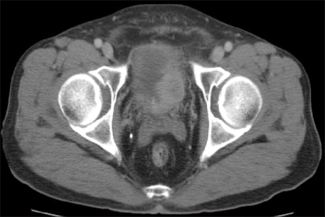
Fig. 1 Multiple irregular marinate massed protruding into the bladder with no lymph node enlargement. The main mass is in the left lateral wall (30×25mm).
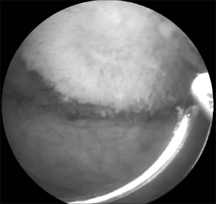
Fig. 2 Cystoscopic finding of a round shape bladder mass in the left lateral wall.

Fig. 3 Light microscopic finding shows evident invasion of small cell carcinoma (arrow) to the muscle layer, which is simulated by lymphocyte-like small round cells (H&E, ×400).
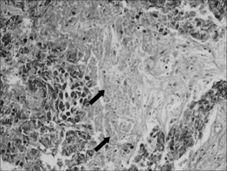
Fig. 4 Necrotic background (arrow) in the tumor cells (H&E, ×400).

Fig. 5 The tumor cells (arrow) show positive immunoreactivity for CD56 (Immunohistochemistry, ×400).
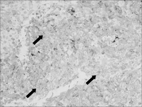
Fig. 6 Immunohistochemical staining with the neuroendocrine marker synaptophysin. Tissues (arrow) are diffusely stained for synaptophysin (Immunohistochemistry, ×400).
Reference
-
1. Mackey JR, Au HJ, Hugh J, Venner P. Genitourinary small cell carcinoma: determination of clinical and therapeutic factors associated with survival. J Urol. 1998. 159:1624–1629.2. Cramer SF, Aikawa M, Cebelin M. Neurosecretory granules in small cell invasive carcinoma of the urinary bladder. Cancer. 1981. 47:724–730.3. Sung DJ, Lee JG, Cho JH. A case of small cell carcinoma of the urinary bladder. Korean J Urol. 1994. 35:1137–1141.4. Lee SK, Moon YT, Kim KD. A case of primary undifferentiated small cell carcinoma of the urinary bladder. Korean J Urol. 1995. 36:454–457.5. Jo SW, Jo IC, Lee OJ, Lee SC, Kim YT, Lee HL, et al. A young man with primary small cell carcinoma of the urinary bladder. Korean J Urol. 2006. 47:556–558.6. Mills SE, Wolfe JT, Weiss MA, Swanson PE, Wick MR, Fowler JE, et al. Small cell undifferentiated carcinoma of the urinary bladder. A light-microscopic, immunocytochemical, and ultrastructural study of 12 cases. Am J Surg Pathol. 1987. 11:606–617.7. Ali SZ, Reuter VE, Zakowski MF. Small cell neuroendocrine carcinoma of the urinary bladder. A clinicopathologic study with emphasis on cytologic features. Cancer. 1997. 79:356–361.8. Grignon DJ, Ro JY, Ayala AG, Shum DT, Ordonez NG, Logothetis CJ, et al. Small cell carcinoma of the urinary bladder. A clinicopathologic analysis of 22 cases. Cancer. 1992. 69:527–536.9. Choong NW, Quevedo JF, Kaur JS. Small cell carcinoma of the urinary bladder. The Mayo Clinic experience. Cancer. 2005. 103:1172–1178.10. Cheng C, Nicholson A, Lowe DG, Kirby RS. Oat cell carcinoma of urinary bladder. Urology. 1992. 38:504–507.
- Full Text Links
-
- Actions
-
Cited
- CITED
-
- Close
- Share
-
- Similar articles
-
- A Case of Small Cell Carcinoma of the Urinary Bladder
- A Case of Primary Undifferentiated Small Cell Carcinoma of the Urinary Bladder
- A Young Man with Primary Small Cell Carcinoma of the Urinary Bladder
- Cytodiagnosis of Primary Small Cell Carcinoma of the Urinary Bladder: A Case Report
- An Elderly Female Non-smoker with Primary Small Cell Carcinoma of the Urinary Bladder
