Evaluation of retrievability using a new soft resin based root canal filling material
- Affiliations
-
- 1Department of Conservative Dentistry, School of Dentistry, Yonsei University, Seoul, Korea. sujungshin@yumc.yonsei.ac.kr
- KMID: 1986875
- DOI: http://doi.org/10.5395/JKACD.2006.31.4.323
Abstract
- The aim of this study was to evaluate the retrievability of Resilon as a root canal filling material. Twenty-seven human single-rooted extracted teeth were instrumented utilizing a crown down technique with Gates-Glidden burs and ProFile system. In group1 (n = 12) canals were obturated with gutta percha and AH-26 plus sealer using a continuous wave technique and backfilled. In group 2 (n = 15) Resilon was used as a filling material. Then teeth were sealed and kept in 37degrees C and 100% humidity for 7 days. For retreatment, the samples were re-accessed and filling material was removed using Gates-Glidden burs and ProFiles. Teeth were sectioned longitudinally to compare the general cleanliness and amount of debris (x 75) using SEM. Chi-square test was used (alpha = 0.05) to analyze the data. The total time required for removal of filling materials was expressed as mean +/- SD (min) and analyzed by the Student t-test (alpha = 0.05). Required time for retreatment was 3.25 +/- 0.32 minutes for gutta percha/AH 26 plus sealer and 3.05 +/- 0.34 minutes for Resilon. There was no statistically significant difference between the two experimental groups. There was no significant difference between the groups in the cleanliness of the root canal wall. This study showed that Resilon was effectively removed by Gates-Glidden burs and ProFiles.
Keyword
MeSH Terms
Figure
-

Figure 1 SEM pictures of sectioned roots after Resilon was removed by ProFiles. Arrows indicate the root canal wall and arrow heads indicate the debris after retreatment. A : Middle portion of root canal wall revealed a clean surface after Resilon filling was removed (score 2). B : Coronal part of root showed multiple sealer debris (score 3). C : Apical root canal wall showed an unclean surface with debris (score 4).
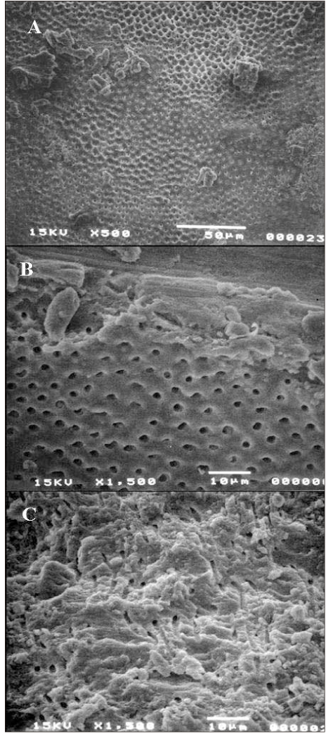
Figure 2 SEM pictures under high magnifications for observation of the patency of dentinal tubules after Resilon removal. Some of specimens showed a clean surface and patent dentinal tubules (A and B). However, in some specimens, the surface was covered with smear layer (C).
Cited by 3 articles
-
Microleakage of resilon: Effects of several self-etching primer
Jong-Hyeon O, Se-Hee Park, Hye-Jin Shin, Kyung-Mo Cho, Jin-Woo Kim
J Korean Acad Conserv Dent. 2008;33(2):133-140. doi: 10.5395/JKACD.2008.33.2.133.Microleakage of resilon by methacrylate-based sealer and self-adhesive resin cement
Sun-Young Ham, Jin-Woo Kim, Hye-Jin Shin, Kyung-Mo Cho, Se-Hee Park
J Korean Acad Conserv Dent. 2008;33(3):204-212. doi: 10.5395/JKACD.2008.33.3.204.Evaluation of softening ability of Xylene & Endosolv-R on three different epoxy resin based sealers within 1 to 2 minutes - an
in vitro study
Pratima Ramakrishna Shenoi, Gautam Pyarelal Badole, Rajiv Tarachand Khode
Restor Dent Endod. 2014;39(1):17-23. doi: 10.5395/rde.2014.39.1.17.
Reference
-
1. Grossman L. Root canal therapy. 1981. Philadelphia: Lea & Febiger;279.2. Gilbert BO Jr, Rice RT. Re-treatment in endodontics. Oral Surg Oral Med Oral Pathol. 1987. 64(3):333–338.
Article3. Baratto Filho F, Ferreira EL, Fariniuk LF. Efficiency of the 0.04 taper ProFile during the re-treatment of gutta-percha-filled root canals. Int Endod J. 2002. 35(8):651–654.
Article4. Barrieshi-Nusair KM. Gutta-percha retreatment: effectiveness of nickel-titanium rotary instruments versus stainless steel hand files. J Endod. 2002. 28(6):454–456.
Article5. Ferreira JJ, Rhodes JS, Ford TR. The efficacy of gutta-percha removal using ProFiles. Int Endod J. 2001. 34(4):267–274.
Article6. Hülsmann M, Bluhm V. Efficacy, cleaning ability and safety of different rotary NiTi instruments in root canal retreatment. Int Endod J. 2004. 37(7):468–476.
Article7. Kosti E, Lambrianidis T, Economides N, Neofitou C. Ex vivo study of the efficacy of H-files and rotary Ni-Ti instruments to remove gutta-percha and four types of sealer. Int Endod J. 2006. 39(1):48–54.
Article8. Masiero AV, Barletta FB. Effectiveness of different techniques for removing gutta-percha during retreatment. Int Endod J. 2005. 38(1):2–7.
Article9. Schirrmeister JF, Wrbas KT, Meyer KM, Altenburger MJ, Hellwig E. Efficacy of different rotary instruments for gutta-percha removal in root canal retreatment. J Endod. 2006. 32(5):469–472.
Article10. Schirrmeister JF, Wrbas KT, Schneider FH, Altenburger MJ, Hellwig E. Effectiveness of a hand file and three nickel-titanium rotary instruments for removing gutta-percha in curved root canals during retreatment. Oral Surg Oral Med Oral Pathol Oral Radiol Endod. 2006. 101(4):542–547.
Article11. Teixeira FB, Teixeira EC, Thompson J, Leinfelder KF, Trope M. Dentinal bonding reaches the root canal system. J Esthet Restor Dent. 2004. 16(6):348–354. discussion 354.
Article12. de Oliveira DP, Barbizam JV, Trope M, Teixeira FB. Comparison between gutta-percha and resilon removal using two different techniques in endodontic retreatment. J Endod. 2006. 32(4):362–364.
Article13. Ezzie E, Fleury A, Solomon E, Spears R, He J. Efficacy of retreatment techniques for a resin-based root canal obturation material. J Endod. 2006. 32(4):341–344.
Article14. Schirrmeister JF, Meyer KM, Hermanns P, Altenburger MJ, Wrbas KT. Effectiveness of hand and rotary instrumentation for removing a new synthetic polymer-based root canal obturation material (Epiphany) during retreatment. Int Endod J. 2006. 39(2):150–156.
Article15. Bramante CM, Betti LV. Efficacy of Quantec rotary instruments for gutta-percha removal. Int Endod J. 2000. 33(5):463–467.
Article
- Full Text Links
-
- Actions
-
Cited
- CITED
-
- Close
- Share
-
- Similar articles
-
- Polymerization shrinkage, hygroscopic expansion and microleakage of resin-based temporary filling materials
- Biocompatibility of root-end filling materials: recent update
- The effects of total-etch, wet-bonding, and light-curing of adhesive on the apical seal of a resin-based root canal filling system
- Micro-computed tomographic evaluation of a new system for root canal filling using calcium silicatebased root canal sealers
- A micro-computed tomographic study of remaining filling materials of two bioceramic sealers and epoxy resin sealer after retreatment
