Korean J Orthod.
2011 Oct;41(5):312-323. 10.4041/kjod.2011.41.5.312.
Comparison of palatal bone thickness between 3D model and lateral cephalometric radiograph
- Affiliations
-
- 1Department of Orthodontics, School of Dentistry, Dankook University, Korea. serail@naver.com
- KMID: 1975385
- DOI: http://doi.org/10.4041/kjod.2011.41.5.312
Abstract
OBJECTIVE
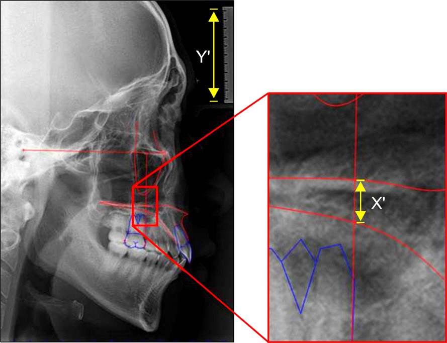
This study compared the bone thickness of the palate between lateral cephalogram and 3D model measurements.
METHODS
The subjects consisted of 30 adults (15 men,15 women) with a normal skeletal pattern and occlusion. The CT images were transformed to a 3D model, and were compared with the cephalometric image. Descriptive statistics for each variable were calculated.
RESULTS
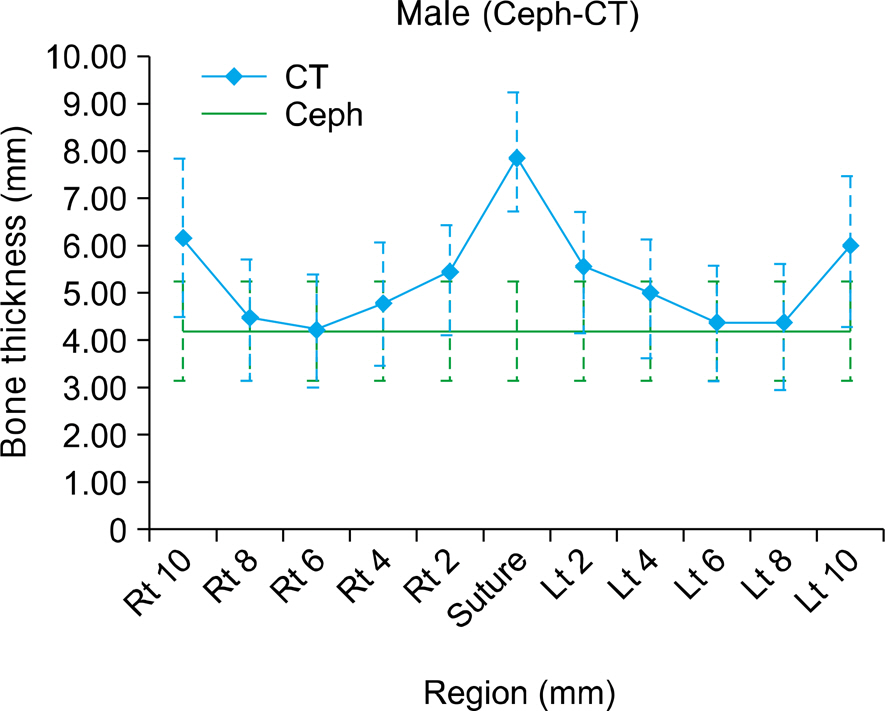
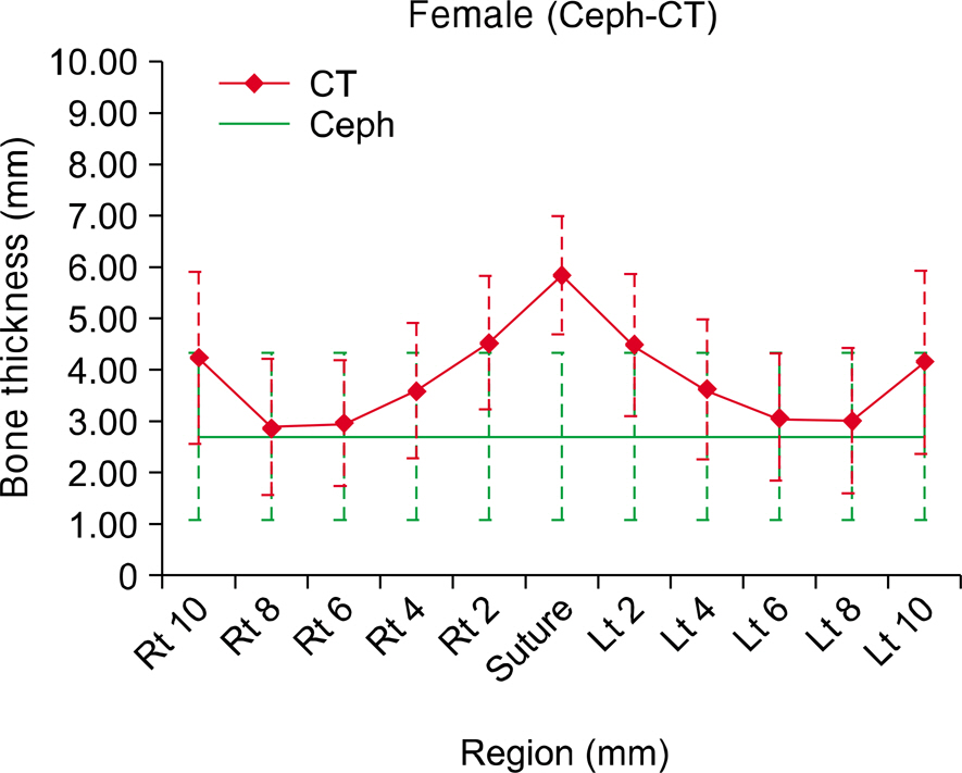
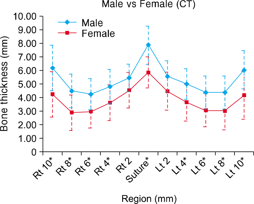
In the 3D CT model, the mid-palatal area was the thickest part. It became thinner as the palate tapered laterally. In the male group, the thinnest portion was positioned 6 mm away from the mid-palate, while in the female group the thinnest portion was 8mm away from the mid-palate. Correlation analysis between the lateral cephalometric and 3D CT model revealed a significant correlation except in the mid palatal area and the area 2 mm lateral to the mid-palate in men, whereas there was a significant relationship in every area in the women. In both men and women, the highest correlation appeared in the area 8 mm lateral to the mid palate.
CONCLUSIONS
Using regression analysis, an actual prediction of the bone thickness between the measured bone thickness of the lateral cephalometric radiograph and 3D model was made. This will provide useful information for mini-implant length selection when inserting into the palate.
Keyword
Figure
Reference
-
1.Kyung SH., Lim JK., Park YC. The use of miniscrew as an anchorage for the orthodontic tooth movement. Korean J Orthod. 2001. 31:415–24.2.Kanomi R. Mini-implant for orthodontic anchorage. J Clin Orthod. 1997. 31:763–7.3.Costa A., Raffainl M., Melsen B. Miniscrews as orthodontic anchorage: a preliminary report. Int J Adult Orthodon Orthognath Surg. 1998. 13:201–9.4.Kyung SH. A study on the bone thickness of midpalatal suture area for miniscrew insertion. Korean J Orthod. 2004. 34:63–70.5.Lim HJ., Eun CS., Cho JH., Lee KH., Hwang HS. Factors associated with initial stability of miniscrews for orthodontic treatment. Am J Orthod Dentofacial Orthop. 2009. 136:236–42.
Article6.Park YC., Lee JS., Kim DH. Anatomical characteristics of the midpalatal suture area for miniscrew implantation using CT image. Korean J Orthod. 2005. 35:35–42.7.Henriksen B., Bavitz B., Kelly B., Harn SD. Evaluation of bone thickness in the anterior hard palate relative to midsagittal orthodontic implants. Int J Oral Maxillofac Implants. 2003. 18:578–81.8.Wehrbein H., Merz BR., Diedrich P. Palatal bone support for orthodontic implant anchorage--a clinical and radiological study. Eur J Orthod. 1999. 21:65–70.9.Kim HJ., Yun HS., Park HD., Kim DH., Park YC. Soft-tissue and cortical-bone thickness at orthodontic implant sites. Am J Orthod Dentofacial Orthop. 2006. 130:177–82.
Article10.Waitzman AA., Posnick JC., Armstrong DC., Pron GE. Craniofacial skeleton measurements based on computed tomography: part I. Accuracy and reproducibility. Cleft Palate Craniofac J. 1992. 29:112–7.11.Masumoto T., Hayashi I., Kawamura A., Tanaka K., Kasai K. Relationships among facial type, buccolingual molar inclination, and cortical bone thickness of the mandible. Eur J Orthod. 2001. 23:15–23.
Article12.Kim DH., Lee JW., Cha KS., Chung DH. Consideration of maxillary sinus bone thickness when installing miniscrews. Korean J Orthod. 2009. 39:354–61.
Article13.Bernhart T., Vollgruber A., Gahleitner A., Dörtbudak O., Haas R. Alternative to the median region of the palate for placement of an orthodontic implant. Clin Oral Implants Res. 2000. 11:595–601.
Article14.Kang S., Lee SJ., Ahn SJ., Heo MS., Kim TW. Bone thickness of the palate for orthodontic mini-implant anchorage in adults. Am J Orthod Dentofacial Orthop. 2007. 131(4 Suppl):S74–81.
Article15.Gracco A., Lombardo L., Cozzani M., Siciliani G. Quantitative cone-beam computed tomography evaluation of palatal bone thickness for orthodontic miniscrew placement. Am J Orthod Dentofacial Orthop. 2008. 134:361–9.
Article16.Baumgaertel S. Quantitative investigation of palatal bone depth and cortical bone thickness for mini-implant placement in adults. Am J Orthod Dentofacial Orthop. 2009. 136:104–8.
Article17.Park HS., Lee YJ., Jeong SH., Kwon TG. Density of the alveolar and basal bones of the maxilla and the mandible. Am J Orthod Dentofacial Orthop. 2008. 133:30–7.
Article18.Park YC., Lee SY., Kim DH., Jee SH. Intrusion of posterior teeth using mini-screw implants. Am J Orthod Dentofacial Orthop. 2003. 123:690–4.
Article19.Kyung SH., Hong SG., Park YC. Distalization of maxillary molars with a midpalatal miniscrew. J Clin Orthod. 2003. 37:22–6.20.Kim SJ., Lim SH. Anatomic study of the incisive canal in relation to midpalatal placement of mini-implant. Korean J Orthod. 2009. 39:146–58.
Article21.Wehrbein H., Merz BR., Hämmerle CH., Lang NP. Bone-to-implant contact of orthodontic implants in humans subjected to horizontal loading. Clin Oral Implants Res. 1998. 9:348–53.22.Motoyoshi M., Inaba M., Ono A., Ueno S., Shimizu N. The effect of cortical bone thickness on the stability of orthodontic mini-implants and on the stress distribution in surrounding bone. Int J Oral Maxillofac Surg. 2009. 38:13–8.
Article23.Harrell WE. 3D orthodontic diagnosis and treatment planning. In: McNamara JA, Kapila S editors. Craniofacial growth series. Vol 43. Digital radiography and three-dimensional imaging. Ann Arbor: The University of Michigan;2006. p. 147–58.
- Full Text Links
- Actions
-
Cited
- CITED
-
- Close
- Share
- Similar articles
-
- 3-Dimensional analysis for class III malocclusion patients with facial asymmetry
- Osteometric Analysis of Palatal Bone Thickness for Orthodontic Miniscrew Placement
- Comparison of measurements from digital cephalometric radiographs and 3D MDCT-synthetized cephalometric radiographs and the effect of head position
- A comparative study between data obtained from conventional lateral cephalometry and reconstructed three-dimensional computed tomography images
- A posteroanterior cephalometric study on the change of maxilla by rapid palatal expansion


