J Korean Assoc Oral Maxillofac Surg.
2014 Jun;40(3):123-129.
A comparative study between data obtained from conventional lateral cephalometry and reconstructed three-dimensional computed tomography images
- Affiliations
-
- 1Department of Oral and Maxillofacial Surgery, Samsung Medical Center, Sungkyunkwan University School of Medicine, Seoul, Korea. hongjr@skku.edu
- 2Department of Nursing, Hanyang University College of Medicine, Seoul, Korea.
Abstract
OBJECTIVES
The aim of this study was to verify the concordance of the measurement values when the same cephalometric analysis method was used for two-dimensional (2D) cephalometric radiography and three-dimensional computed tomography (3D CT), and to identify which 3D Frankfort horizontal (FH) plane was the most concordant with FH plane used for cephalometric radiography.
MATERIALS AND METHODS
Reference horizontal plane was FH plane. Palatal angle and occlusal plane angle was evaluated with FH plane. Gonial angle (GA), palatal angle, upper occlusal plane angle (UOPA), mandibular plane angle (MPA), U1 to occlusal plane angle, U1 to FH plane angle, SNA and SNB were obtained on 2D cephalmetries and reconstructed 3D CT. The values measured eight angles in 2D lateral cephalometry and reconstructed 3D CT were evaluated by intraclass correlation coefficiency (ICC). It also was evaluated to identify 3D FH plane with high degree of concordance to 2D one by studying which one in four FH planes shows the highest degree of concordance with 2D FH plane.
RESULTS
ICCs of MPA (0.752), UOPA (0.745), SNA (0.798) and SNB (0.869) were high. On the other hand, ICCs of gonial angle (0.583), palatal angle (0.287), U1 to occlusal plane (0.404), U1 to FH plane (0.617) were low respectively. Additionally GA and MPA acquired from 2D were bigger than those on 3D in all 20 patients included in this study. Concordance between one UOPA from 2D and four UOPAs from 3D CT were evaluated by ICC values. Results showed no significant difference among four FH planes defined on 3D CT.
CONCLUSION
FH plane that can be set on 3D CT does not have difference in concordance from FH plane on lateral cephalometry. However, it is desirable to define FH plane on 3D CT with two orbitales and one porion considering the reproduction of orbitale itself.
Figure
-
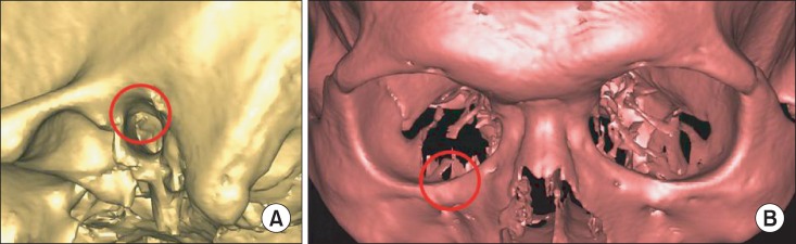
Fig. 1 A. Large, round-shaped bony edge around the porion. B. Sharp edge around the orbitale.
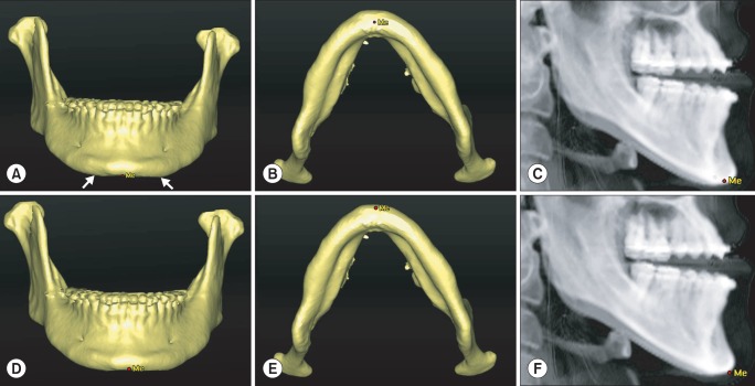
Fig. 2 A. Downward extended adjacent structure on both sides (white arrows). B. Position of the menton (Me) in the inferior-superior direction. C. Position of the Me on three-dimensional (3D) cephalometry. D. Me positioned in the anterior-posterior view. E. Me positioned in the anterior view. F. Position of the Me on 3D cephalometry.
Reference
-
1. van Vlijmen OJ, Bergé SJ, Swennen GR, Bronkhorst EM, Katsaros C, Kuijpers-Jagtman AM. Comparison of cephalometric radiographs obtained from cone-beam computed tomography scans and conventional radiographs. J Oral Maxillofac Surg. 2009; 67:92–97. PMID: 19070753.
Article2. Athanasiou AE. Orthodontic cephalometry. London: Mosby-Wolfe;1997.3. Adams GL, Gansky SA, Miller AJ, Harrell WE Jr, Hatcher DC. Comparison between traditional 2-dimensional cephalometry and a 3-dimensional approach on human dry skulls. Am J Orthod Dentofacial Orthop. 2004; 126:397–409. PMID: 15470343.
Article4. Miller PA, Savara BS, Singh IJ. Analysis of errors in cephalometric measurement of three-dimensional distances on the maxilla. Angle Orthod. 1966; 36:169–175. PMID: 5218677.5. Gravely JF, Benzies PM. The clinical significance of tracing error in cephalometry. Br J Orthod. 1974; 1:95–101. PMID: 4525738.
Article6. Midtgård J, Björk G, Linder-Aronson S. Reproducibility of cephalometric landmarks and errors of measurements of cephalometric cranial distances. Angle Orthod. 1974; 44:56–61. PMID: 4520951.7. Trpkova B, Major P, Prasad N, Nebbe B. Cephalometric landmarks identification and reproducibility: a meta analysis. Am J Orthod Dentofacial Orthop. 1997; 112:165–170. PMID: 9267228.
Article8. Wylie GA, Fish LC, Epker BN. Cephalometrics: a comparison of five analyses currently used in the diagnosis of dentofacial deformities. Int J Adult Orthodon Orthognath Surg. 1987; 2:15–36. PMID: 3469281.9. Magalhaes AE, Stella JP, Epker BN. Facial anthropometrics versus cephalometry as predictors for surgical treatment in patients with class III dentofacial deformities. Int J Adult Orthodon Orthognath Surg. 1995; 10:295–302. PMID: 9082019.10. Swennen GR, Schutyser F. Three-dimensional virtual approach to diagnosis and treatment planning of maxillo-facial deformity. In : Bell W, editor. Distraction osteogenesis of the facial skeleton. Hamilton, ON: BC Decker Inc;2007. p. 55.11. Maki K, Okano T, Morohashi T, Yamada S, Shibaski Y. The application of three-dimensional quantitative computed tomography to the maxillofacial skeleton. Dentomaxillofac Radiol. 1997; 26:39–44. PMID: 9446989.
Article12. Vannier MW, Marsh JL, Warren JO. Three dimensional CT reconstruction images for craniofacial surgical planning and evaluation. Radiology. 1984; 150:179–184. PMID: 6689758.
Article13. Trpkova B, Prasad NG, Lam EW, Raboud D, Glover KE, Major PW. Assessment of facial asymmetries from posteroanterior cephalograms: validity of reference lines. Am J Orthod Dentofacial Orthop. 2003; 123:512–520. PMID: 12750669.
Article14. Wong RW, Chau AC, Hägg U. 3D CBCT McNamara's cephalometric analysis in an adult southern Chinese population. Int J Oral Maxillofac Surg. 2011; 40:920–925. PMID: 21511439.
Article15. Maeda M, Katsumata A, Ariji Y, Muramatsu A, Yoshida K, Goto S, et al. 3D-CT evaluation of facial asymmetry in patients with maxillofacial deformities. Oral Surg Oral Med Oral Pathol Oral Radiol Endod. 2006; 102:382–390. PMID: 16920547.
Article16. Chebib FS, Chamma AM. Indices of craniofacial asymmetry. Angle Orthod. 1981; 51:214–226. PMID: 6943950.17. Epker BN, Fish LC, Stella JP. Dentofacial deformities: integrated orthodontic and surgical correction. St Louis: CV Mosby;1998. p. 29–33.18. Raustia AM, Salonen MA. Gonial angles and condylar and ramus height of the mandible in complete denture wearers--a panoramic radiograph study. J Oral Rehabil. 1997; 24:512–516. PMID: 9250838.
Article19. Piedra I. The Levandoski panoramic analysis in the diagnosis of facial and dental asymmetries. J Clin Pediatr Dent. 1995; 20:15–21. PMID: 8634190.20. Mattila M, Könönen M, Mattila K. Vertical asymmetry of the mandibular ramus and condylar heights measured with a new method from dental panoramic radiographs in patients with psoriatic arthritis. J Oral Rehabil. 1995; 22:741–745. PMID: 8606331.
Article21. Updegrave WJ. Visualizing the mandibular ramus in panoramic radiography. Oral Surg Oral Med Oral Pathol. 1971; 31:422–429. PMID: 5277395.
Article22. Yeo DK, Freer TJ, Brockhurst PJ. Distortions in panoramic radiographs. Aust Orthod J. 2002; 18:92–98. PMID: 12462686.23. Cavalcanti MG, Haller JW, Vannier MW. Three-dimensional computed tomography landmark measurement in craniofacial surgical planning: experimental validation in vitro. J Oral Maxillofac Surg. 1999; 57:690–694. PMID: 10368094.
Article24. Hildebolt CF, Vannier MW, Knapp RH. Validation study of skull three-dimensional computerized tomography measurements. Am J Phys Anthropol. 1990; 82:283–294. PMID: 2375381.
Article25. Mulick JF. Clinical use of the frontal headfilm. Angle Orthod. 1965; 35:299–304. PMID: 5213520.
- Full Text Links
-
- Actions
-
Cited
- CITED
-
- Close
- Share
-
- Similar articles
-
- Comparative study of glenoid version and inclination using two-dimensional images from computed tomography and three-dimensional reconstructed bone models
- Comparison Analysis of 3D CT and Cephalometrics in Craniofacial Measurements
- Accuracy of three-dimensional cephalograms generated using a biplanar imaging system
- Comparison of conventional lateral cephalograms with corresponding CBCT radiographs
- Three-dimensional image analysis of the skull using variable CT scanning protocols-effect of slice thickness on measurement in the three-dimensional CT images
