3-Dimensional analysis for class III malocclusion patients with facial asymmetry
- Affiliations
-
- 1Department of Oral and Maxillofacial Surgery, Wonkwang University Dental Hospital, Iksan, Korea. mistej@naver.com
- 2Wonkwang University Dental Research Institute, Iksan, Korea.
Abstract
OBJECTIVES
The aim of this study is to investigate the correlation between 2-dimensional (2D) cephalometric measurement and 3-dimensional (3D) cone beam computed tomography (CBCT) measurement, and to evaluate the availability of 3D analysis for asymmetry patients.
MATERIALS AND METHODS
A total of Twenty-seven patients were evaluated for facial asymmetry by photograph and cephalometric radiograph, and CBCT. The 14 measurements values were evaluated and those for 2D and 3D were compared. The patients were classified into two groups. Patients in group 1 were evaluated for symmetry in the middle 1/3 of the face and asymmetry in the lower 1/3 of the face, and those in group 2 for asymmetry of both the middle and lower 1/3 of the face.
RESULTS
In group 1, significant differences were observed in nine values out of 14 values. Values included three from anteroposterior cephalometric radiograph measurement values (cant and both body height) and six from lateral cephalometric radiographs (both ramus length, both lateral ramal inclination, and both gonial angles). In group 2, comparison between 2D and 3D showed significant difference in 10 factors. Values included four from anteroposterior cephalometric radiograph measurement values (both maxillary height, both body height) and six from lateral cephalometric radiographs (both ramus length, both lateral ramal inclination, and both gonial angles).
CONCLUSION
Information from 2D analysis was inaccurate in several measurements. Therefore, in asymmetry patients, 3D analysis is useful in diagnosis of asymmetry.
Figure
-
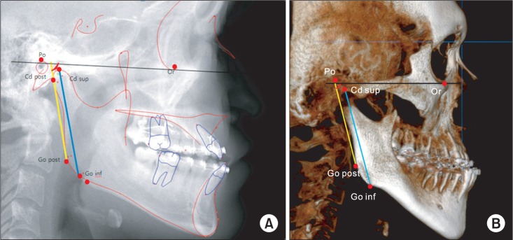
Fig. 1 The measurement of lateral cephalogrphic radiography with V-ceph program (A) and 3-dimensional image with OnDemand program (B). In 2-dimensional image, the program divided right and left side based on the fact that closer part to film tends to show more radiopaque in lateral cephalogram. Lateral ramal inclination was represented the red angle between posterior ramal border (yellow line) and Frankfort horizontal plane (black line). Ramal length displayed blue line, and we measured gonial angle formed by Ar-Go-Me. (Ar: articulare, Go: gonion, Me: menton, Po: porion, Cd post: posterior condyle point, Cd sup: highest point of the condyle, Go post: posterior gonion point, Go inf: lowest point of the Go area, Or: orbitale)
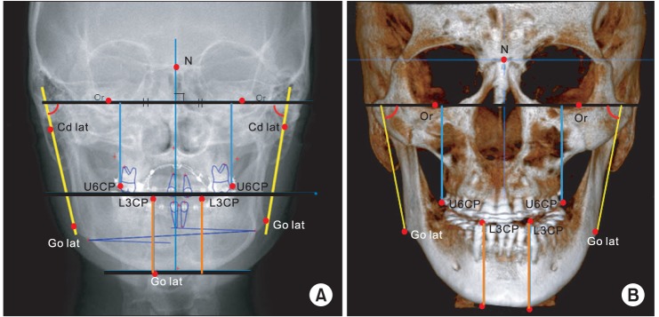
Fig. 2 The measurement of antero-posterior cephalographic radiograph. The line connecting both orbitale was used as a standard. Midsagittal line perpendicular to the line connecting both orbitale and passing nasion point was used as a vertical standard (A). The measurement of 3-dimensional image with OnDemand program. The plane including both orbitale and porion was a standard plane and the plane passing nasion and dens is midsagittal plane (B). Frontal ramal inclination (red angle): angle between both orbitale line (black line) and yellow line from lateral condylar point and lateral Go point, body height: orange line, maxillary height: blue line. (N: nasion, Or: orbitale, Cd lat: lateral condyle point, U6CP: upper first molar buccomesial cusp point, L3CP: mandible canine cusp point, Go lat: lateral gonion point)
Reference
-
1. Fukushima K, Yasui K, Oatuka Y, Matsui S, Hirase N, Takayanagi J, et al. Morphological characteristics of patients with jaw deformity -Frontal cepharometric evaluation of facial symmetry-. Meikai Univ Dent J. 2003; 32:118–123.2. Tani M, Iketani M, Watanabe M, Suda S, Fujimura N, Miyazawa M, et al. Posterior-anterior cephalometric analysis in patients with dentofacial deformities. J Jpn Stomatol Soc. 1989; 35:1749–1759.
Article3. Severt TR, Proffit WR. The prevalence of facial asymmetry in the dentofacial deformities population at the University of North Carolina. Int J Adult Orthodon Orthognath Surg. 1997; 12:171–176. PMID: 9511487.4. Samman N, Tong AC, Cheung DL, Tideman H. Analysis of 300 dentofacial deformities in Hong Kong. Int J Adult Orthodon Orthognath Surg. 1992; 7:181–185. PMID: 1291612.5. Sassouni V. Diagnosis and treatment planning via roentgenographic cephalometry. Am J Orthodontics. 1958; 44:433–463.
Article6. Ricketts RM. Cephalometric synthesis. Am J Orthod. 1960; 46:647–673.
Article7. Mulick JF. Clinical use of the frontal headfilm. Angle Orthod. 1965; 35:299–304. PMID: 5213520.8. Graber TM. New horizons in case analysis-clinical cephalometrics. Am J Orthodont. 1952; 38:603–624.
Article9. Graber TM. A critical review of clinical cephalometric radiography. Am J Orthodont. 1954; 40:1–26.
Article10. Keen RR, Callahan GR. Osteochondroma of the mandibular condyle: report of case. J Oral Surg. 1977; 35:140–143. PMID: 264508.11. Gateno J, Xia JJ, Teichgraeber JF. Effect of facial asymmetry on 2-dimensional and 3-dimensional cephalometric measurements. J Oral Maxillofac Surg. 2011; 69:655–662. PMID: 21353927.
Article12. Trpkova B, Prasad NG, Lam EW, Raboud D, Glover KE, Major PW. Assessment of facial asymmetries from posteroanterior cephalograms: validity of reference lines. Am J Orthod Dentofacial Orthop. 2003; 123:512–520. PMID: 12750669.
Article13. Kawamata A, Ariji Y, Langlais RP. Three-dimensional computed tomography imaging in dentistry. Dent Clin North Am. 2000; 44:395–410. PMID: 10740775.14. Kawamata A, Fujishita M, Ariji Y, Ariji E. Three-dimensional computed tomographic evaluation of morphologic airway changes after mandibular setback osteotomy for prognathism. Oral Surg Oral Med Oral Pathol Oral Radiol Endod. 2000; 89:278–287. PMID: 10710450.
Article15. Haraguchi S, Takada K, Yasuda Y. Facial asymmetry in subjects with skeletal Class III deformity. Angle Orthod. 2002; 72:28–35. PMID: 11843270.16. Cheney EA. Dentofacial asymmetries and their clinical significance. Am J Orthod. 1961; 47:814–829.
Article17. Greiner M, Greiner A, Hirschfelder U. Variance of landmarks in digital evaluations: comparison between CT-based and conventional digital lateral cephalometric radiographs. J Orofac Orthop. 2007; 68:290–298. PMID: 17639277.
Article18. Silling G, Rauch MA, Pentel L, Garfinkel L, Halberstadt G. The significance of cephalometrics in treatment planning. Angle Orthod. 1979; 49:259–262. PMID: 292348.19. Nijkamp PG, Habets LL, Aartman IH, Zentner A. The influence of cephalometrics on orthodontic treatment planning. Eur J Orthod. 2008; 30:630–635. PMID: 18981169.
Article20. Turpin DL. British Orthodontic Society revises guidelines for clinical radiography. Am J Orthod Dentofacial Orthop. 2008; 134:597–598. PMID: 18984385.
Article21. Houston WJ. The analysis of errors in orthodontic measurements. Am J Orthod. 1983; 83:382–390. PMID: 6573846.
Article22. van Vlijmen OJ, Maal TJ, Bergé SJ, Bronkhorst EM, Katsaros C, Kuijpers-Jagtman AM. A comparison between two-dimensional and three-dimensional cephalometry on frontal radiographs and on cone beam computed tomography scans of human skulls. Eur J Oral Sci. 2009; 117:300–305. PMID: 19583759.
Article23. van Vlijmen OJ, Bergé SJ, Swennen GR, Bronkhorst EM, Katsaros C, Kuijpers-Jagtman AM. Comparison of cephalometric radiographs obtained from cone-beam computed tomography scans and conventional radiographs. J Oral Maxillofac Surg. 2009; 67:92–97. PMID: 19070753.
Article24. Yitschaky O, Redlich M, Abed Y, Faerman M, Casap N, Hiller N. Comparison of common hard tissue cephalometric measurements between computed tomography 3D reconstruction and conventional 2D cephalometric images. Angle Orthod. 2011; 81:11–16. PMID: 20936949.
Article25. Hwang HS, Hwang CH, Lee KH, Kang BC. Maxillofacial 3-dimensional image analysis for the diagnosis of facial asymmetry. Am J Orthod Dentofacial Orthop. 2006; 130:779–785. PMID: 17169741.
Article26. Gateno J, Xia JJ, Teichgraeber JF. New 3-dimensional cephalometric analysis for orthognathic surgery. J Oral Maxillofac Surg. 2011; 69:606–622. PMID: 21257250.
Article27. Damstra J, Fourie Z, Ren Y. Evaluation and comparison of postero-anterior cephalograms and cone-beam computed tomography images for the detection of mandibular asymmetry. Eur J Orthod. 2013; 35:45–50. PMID: 21459833.
Article28. Yoon YJ, Kim KS, Hwang MS, Kim HJ, Choi EH, Kim KW. Effect of head rotation on lateral cephalometric radiographs. Angle Orthod. 2001; 71:396–403. PMID: 11605875.
- Full Text Links
-
- Actions
-
Cited
- CITED
-
- Close
- Share
-
- Similar articles
-
- A cehalometric study on facial morphology in angle's Class III malocclusion patients with facial asymmetry
- The attrition pattern in Angle Class III malocclusion with facial asymmetry
- Cone-beam computed tomography analysis of transverse dental compensation in patients with skeletal Class III malocclusion and facial asymmetry
- Comparison of changes in the transverse dental axis between patients with skeletal Class III malocclusion and facial asymmetry treated by orthognathic surgery with and without presurgical orthodontic treatment
- A comparative study of mandibular tooth development between Angle Class I malocclusion group and Angle Class III malocclusion group
