Minimally Invasive Plate Osteosynthesis to Prevent or Treat the Deformity after Distraction Osteogenesis
- Affiliations
-
- 1Department of Orthopedic Surgery, College of Medicine, Kyungpook National University, Daegu, Korea. cwoh@knu.ac.kr
- KMID: 1947783
- DOI: http://doi.org/10.4055/jkoa.2007.42.6.764
Abstract
-
PURPOSE: To determine the efficacy of minimally invasive plate osteosynthesis (MIPO) for distraction callus after a lengthening procedure with an external fixator.
MATERIALS AND METHODS
Distraction osteogenesis were performed using an external fixator for growth arrest (7 cases), congenital pseudoarthrosis of tibia (2 cases), and congenital leg length discrepancy (2 cases). MIPO was performed using a locking compression plate over the distraction callus. The mean age of the index procedure was 11 years, and mean amount of distraction was 5.4 cm. Eight were treated earlier to remove the external fixator after achieving of the target length, and 3 were treated as a salvage operation of fractures after removal.
RESULTS
In all patients, the distraction callus healed with its length or correction into the original alignment maintained. The mean external fixation index was 34.3 days/cm and the mean healing index was 52.6 days/cm. In 8 patients with the early removal of the external fixator, the mean external fixation index was 26.9 days/cm. No patient developed a deep infection or implant failure. All patients recovered their preoperative joint motion and were satisfied with their function.
CONCLUSION
MIPO can prevent or correct a deformity after distraction osteogenesis, and allow patients to return to their daily life earlier.
MeSH Terms
Figure
-

Fig. 1 (Case No. 4) A 9 year-old girl with post-septic growth arrest underwent tibial lengthening with ilizarov ring fixator (A & B). After achieving desired length (C), percutaneous plating was performed using two plates in view of small size of tibia (D). Fixator was removed and alignment is well maintained (E). External fixation index was 15.1 days/cm, and healing index was 45.6days/cm with the good consolidation of callus (F). The patient suffered a superficial infection. But, she could have a good function after removal of plates (G & H).
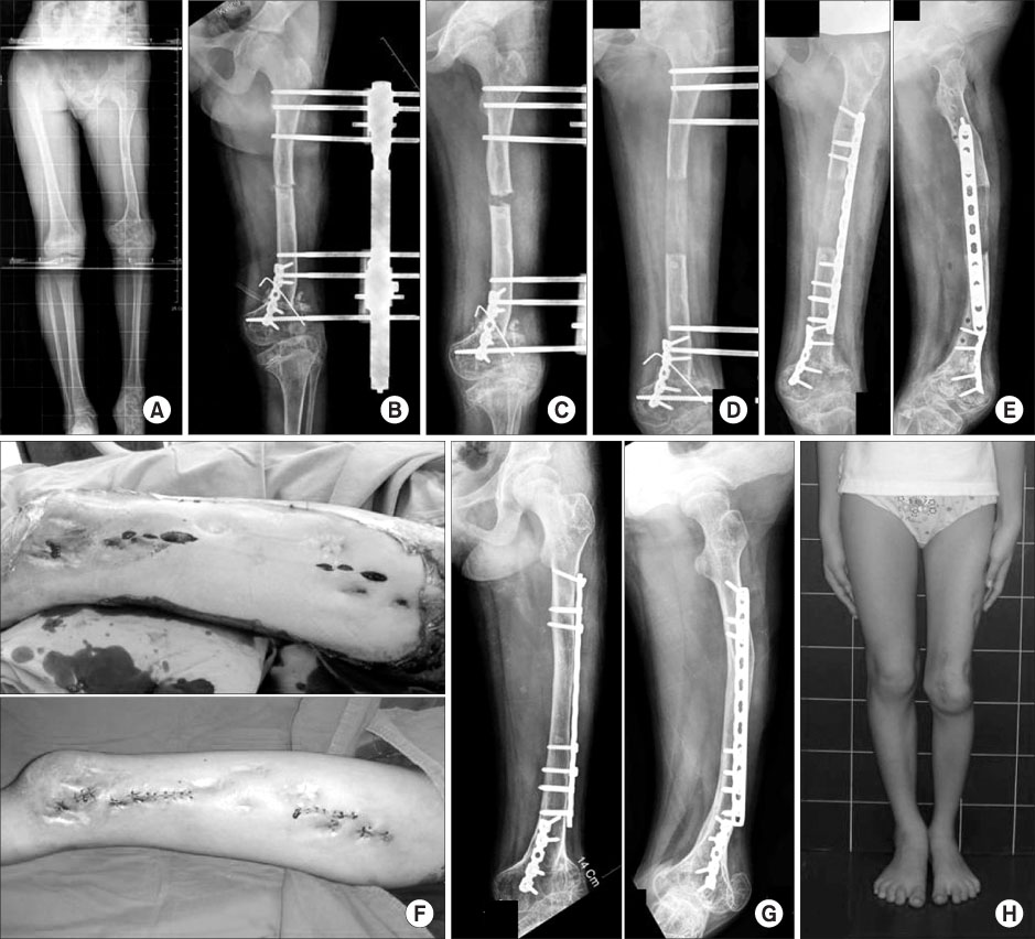
Fig. 2 (Case No. 5) A 10 year-old girl had a leg length discrepancy with deformity due to traumatic growth arrest of the distal femur and proximal tibia. With corrective osteotomy of the distal femur, femoral lengthening was conducted with a monolateral external fixator (A & B). After achieving target length, percutaneous plating was done under the fluoroscopic guide (C & D). The distraction segment was well maintained (E), after the external fixator was removed (F). At 14 months postoperatively, the distraction callus healed (G), and the patient gained the equalization of legs (H).

Fig. 3 (Case No. 9). A 5 year-old girl with femoral shortening (A) underwent limb lengthening with monolateral fixtor (B&C). With maturing of the distraction callus, the external fixtor was removed (D). But, a varus collapse occurred at 1 week after removal (E). Percutaneous plating was done with correction of deformed callus (F). Good alignment was achieved after operation (G). Although a small plate was fixed for stress fracture at the previous pin track, the distraction callus matured with marrow canalization (H) and she showed an excellent function (I).
Reference
-
1. Baumgart R, Betz A, Schweiberer L. A fully implantable motorized intramedullary nail for limb lengthening and bone transport. Clin Orthop Relat Res. 1997. 343:135–143.
Article2. Cattaneo R, Catagni M, Johnson EE. The treatment of infected nonunions and segmental defects of the tibia by the methods of Ilizarov. Clin Orthop Relat Res. 1992. 280:143–152.
Article3. Garcia-Cimbrelo E, Curto de la Mano A, Garcia-Rey E, Cordero J, Marti-Ciruelos R. The intramedullary elongation nail for femoral lengthening. J Bone Joint Surg Br. 2002. 84:971–977.4. Hahn SB, Park HW, Kang KW. Limb lengthening by gradual elongation intramedullary nail (Albizzia). J Korean Orthop Assoc. 1998. 33:343–349.5. Herzenberg JE, Scheufele PT, Paley D, Bechtel R, Tepper S. Knee range of motion in isolated femoral lengthening. Clin Orthop Relat Res. 1994. 301:49–54.
Article6. Hofmann GO, Gonschorek O, Bühren V. Segment transport employing intramedullary devices in tibial bone defects following trauma and infection. J Orthop Trauma. 1999. 13:170–177.
Article7. Ilizarov GA. Clinical application of the tension-stress effect for limb lengthening. Clin Orthop Relat Res. 1990. 250:8–26.
Article8. Kristiansen LP, Steen H. Lengthening of tibia over an intramedullary nail, using the Ilizarov external fixator: major complications and slow consolidation of 9 lengthenings. Acta Orthop Scand. 1990. 70:271–274.9. Marsh JL, Prokuski , Biermann JS. Chronic infected tibial nonunions with bone loss: conventional techniques versus bone transport. Clin Orthop Relat Res. 1994. 301:139–146.10. Mekhail AO, Abraham E, Gruber B, Gonzalez M. Bone transport in the management of posttraumatic bone defects in the lower extremity. J Trauma. 2004. 56:368–378.
Article11. Oh CW, Min WK, Kyung HS, et al. Internal bone transport in the management of tibial bone defects. J Korean Fracture Soc. 2005. 18:36–42.
Article12. Oh CW, Oh JK, Kyung HS, et al. Double plating of unstable proximal tibial fractures using minimally invasive percutaneous osteosynthesis technique. Acta Orthop. 2006. 77:524–530.
Article13. Oh CW, Kyung HS, Park IH, Kim PT, Ihn JC. Distal tibia metaphyseal fractures treated by percutaneous plate osteosynthesis. Clin Orthop Relat Res. 2003. 408:286–291.
Article14. Paley D. Problems, obstacles, and complications of limb lengthening by the Ilizarov technique. Clin Orthop Relat Res. 1990. 250:81–104.
Article15. Paley D, Herzenberg JE, Paremain G, Bhave A. Femoral lengthening over an intramedullary nail: a matched-case comparison with Ilizarov lengthening. J Bone Joint Surg Am. 1997. 79:1464–1480.16. Raschke MJ, Mann JW, Oedekoven G, Claudi BF. Segmental transport after unreamed intramedullary nailing: preliminary report of a "Monorail" system. Clin Orthop Relat Res. 1992. 282:233–240.17. Singh S, Lahiri A, Iqbal M. The results of limb lengthening by callus distraction using an extending intramedullary nail (Fitbone) in non-traumatic disorders. J Bone Joint Surg Br. 2006. 88:938–942.
Article18. Song HR, Oh CW, Mattoo R, et al. Femoral lengthening with external fixator over the intramedullary nail. J Korean Orthop Assoc. 2004. 39:812–818.
Article19. Stoffel K, Dieter U, Stachowiak G, Gachter A, Kuster MS. Biomechanical testing of the LCP--how can stability in locked internal fixators be controlled? Injury. 2003. 34:Suppl 2. B11–B19.
- Full Text Links
-
- Actions
-
Cited
- CITED
-
- Close
- Share
-
- Similar articles
-
- Distraction Osteogenesis Combined with a Plate to Treat Brachymetacarpia
- Clinical Outcomes of Locking Compression Plate Fixation through Minimally Invasive Percutaneous Plate Osteosynthesis in the Treatment of Distal Tibia Fracture
- Distraction Osteogenesis Update: Introduction of Multidirectional Cranial Distraction Osteogenesis
- Minimally Invasive Percutaneous Plate Osteosynthesis Using Periarticular Plate for Distal Tibial Fractures
- Minimally Invasive Percutaneous Plate Stabilization Using a Medial Locking Plate for Proximal Tibial Fractures: Technical Note
