J Korean Soc Spine Surg.
2007 Sep;14(3):129-136. 10.4184/jkss.2007.14.3.129.
Radiographic Morphometry of Lumbar Intervertebral Disc Space in Normal Korean
- Affiliations
-
- 1Department of Orthopedic Surgery, Yonsei University College of Medicine. shmoon@yuhs.ac
- 2Department of Orhtopaedic Surgery, Korea University College of Medicine.
- KMID: 1941643
- DOI: http://doi.org/10.4184/jkss.2007.14.3.129
Abstract
-
STUDY DESIGN: Radiographic measurement of the lumbar disc height
OBJECTIVES
To measure the lumbar disc height on the radiographs in normal Koreans. SUMMARY OF LITERATURE REVIEW: Many reports show good results after many procedures, such as inter-vertebral body fusion using a cage or total disc replacement, which restores the adequate disc height. However, there are no references regarding the range of normal lumbar disc heights in Korean adults, which can be used as a standard for the implant size.
MATERIALS AND METHODS
One hundred and thirty two subjects (age range 20 to 40 years), who had no previous history of low back pain and no significant findings on the physical examination, were enrolled in this study. The plain lateral lumbar spine radiograph was taken in the supine position. The intervertebral disc heights were measured at the anterior, middle and posterior portion of each lumbar disc. The average magnification rate was 115%, and the disc heights were corrected by the magnification rate in each segment.
RESULTS
The lumbar disc height showed a cranio-caudal pattern in both the male and female groups. The L4-5 disc heights were highest at the anterior, middle and posterior portion in males. The L4-5 disc heights were highest at the middle and posterior portion in females. The L5-S1 disc height was highest at the anterior portion in females, but there was no significant difference between the L4-5 and L5-S1 disc height at the anterior portion. There was no significant difference in disc height between males and females except at the anterior portion of the L1-2 and L2-3 disc. There was no significant decrease in disc height in overweight people at all measured site in males and females except at the posterior portion of the L1-2 disc in males.
CONCLUSION
This study is meaningful in that it provides a reference value for the lumbar disc height in Korean adults. The measured values might also be useful for manufacturing a Korean model of an artificial lumbar disc prosthesis or surgical instruments for lumbar interbody fusion.
Keyword
MeSH Terms
Figure
-
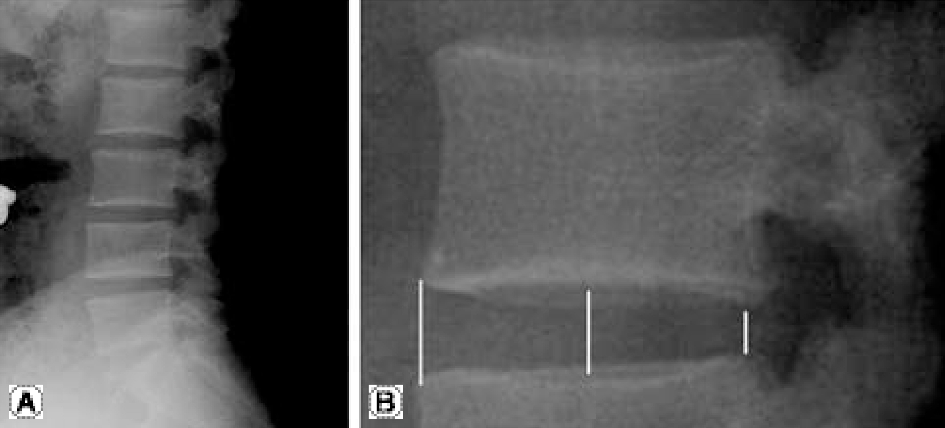
Fig. 1. Measuring method for anterior, middle and posterior portion of disc height. (A) lateral plain film for lumbar spine centerd on L4 vertebral body. (B) After 4 fold magnification, anterior, middle and posterior portion of disc height were measured.

Fig. 2. Male disc height (mm) for anterior, middle and posterior portion.

Fig. 3. Female disc height (mm) for anterior, middle and posterior portion.
Reference
-
1). Matsumura A, Taneichi H, Suda K, Kajino T, Moridaira H, Kaneda K. Comparative study of radiographic disc height changes using two different interbody devices for transforaminal lumbar interbody fusion: open box vs. fenestrated tube interbody cage. Spine. 2006; 31:E871–876.2). Bertagnoli R, Yue JJ, Kershaw T, et al. Lumbar total disc arthroplasty utilizing the ProDisc prosthesis in smok-ers versus nonsmokers: a prospective study with 2-year minimum followup. Spine. 2006; 31:992–997.3). Kim NH, Lee HM, Chung IH, Kim HJ, Kim SJ. Morphometric study of the pedicles of thoracic and lumbar vertebrae in Koreans. Spine. 1994; 19:1390–1394.
Article4). Kim NH, Moon SH, Lee HM, Kim DH. Spinal Dimensions and Shape Variation in Koreans - Radiographic Quantitative Morphometry. J Korean Orthop Assoc. 1998; 33:1611–1619.5). Kimura S, Steinmach GC, Watenpaugh DE, Gargens AR. Lumbar spine disc height and curvature responses to an axial load generated by a compression device compati-ble with magnetic resonance imaging. Spine. 2001; 26:2596–2600.
Article6). Keller TS, Nathan M. Height change caused by creep in intervertebral discs: a sagittal plane model. J Spinal Disord. 1999; 12:313–324.7). Farfan HF. Mechanical disorders of the low back. Lea and Febiger. Philadelphia 1973. (cited from Frobin W, Brinckmann P, Biggemann M, Tillotson M, Burton K: Precision measurement of disc height, vertebral height and sagittal plane displacement from lateral radiographic views of the lumbar spine. Clin Biomech. 1997; 12:1–63. .).8). Hurxthal LM. Measurement of anterior vertebral compressions and biconcave vertebrae. J Am Radiol. 1968; 103:635–644.
Article9). 4Frobin W, Brinckmann P, Biggemann M, Tillotson M, Burton K. Precision measurement of disc height, vertebral height and sagittal plane displacement from lateral radiographic views of the lumbar spine. Clin Biomech. 1997; 12:1–63.10). Luoma K, Riihimaki H, Luukkonen R, Raininko R, Viikari-Juntura E, Lamminen A. Low back pain in relation to lumbar disc degeneration. Spine. 2000; 25:487–492.
Article11). Riihimaki H, Viikari-Juntura E. Back and limb disorders (in McDonald C, Wheatley M eds. Epidemiology of Work Related Diseases. London: BMJ Book;p. 223–243. 2000.12). Kraemer J, Kolditz D, Gowin R. Water and electrolyte content of human intervertebral discs under variable load. Spine. 1985; 10:69–71.
Article13). Butler D, Trafimow JH, Andersson GB, McNeill TW, Huckman MS. Discs degenerate before facets. Spine. 1990; 15:111–113.
Article14). Lemaire JP, Carrier H, Ali el-HS, Skalli W, Lavaste F. Clinical and radiological outcomes with the Charite artificial disc: a 10-year minimum followup. J Spinal Disord Tech. 2005; 18:353–359.15). Mayer HM, Korge A. Non-fusion technology in degenerative lumbar spinal disorders: Facts, questions, chal-lenges. Eur Spine J. 2002; 11:111–114.
Article16). Gamradt SC, Wang JC. Contemporary concepts review: Lumbar disc arthroplasty. Spine J. 2005; 5:95–103.17). Rohlmann A, Zander T, Bergmann G. Effect of total disc replacement with proDisc on intersegmental rotation of the lumbar. Spine. 2005; 30:738–743.18). Frobin W, Brinckmann P, Biggemann M. Objektive messung der Hohe lumbaler bandscheiben aus seitlichen Rontgen-ubersichtsaufnahmen. Z orthop. 1997; 135:394–402. (cited from Shao Z, Rompe G, Schiltenwolf M: Radiographic changes in the lumbar intervertebral discs and lumbar vertebrae with age. Spine. 2002; 27:163–168. .).19). Riihimaki H, Mattsson T, Zitting A, Wickstrom G, Hanninen K, Waris P. Radiographically detectable degenerative changes of the lumbar spine among concrete reinforcement workers and house painters. Spine. 1990; 15:114–119.
Article20). Parkkola R, Rytokoski U, Kormano M. Magnetic resonance imaging of the discs and trunk muscles in patients with chronic low back pain and healthy control subjects. Spine. 1993; 18:830–836.
Article21). Elfering A, Semmer N, Birkhofer D, Zanetti M, Hodler J, Boos N. Risk factors for lumbar disc degeneration: a 5-year prospective MRI study in asymptomatic individuals. Spine. 2002; 27:125–134.22). Bostman OM. Prevalence of obesity among patients admitted for elective orthopaedic surgery. Int J Obes Relat Metab Disord. 1994; 18:709–713.23). Liuke M, Solovieva S, Lamminen A, et al. Disc degeneration of the lumbar spine in relation to overweight. Int J Obes (Lond). 2005; 29:903–908.
Article
- Full Text Links
-
- Actions
-
Cited
- CITED
-
- Close
- Share
-
- Similar articles
-
- The Change of Intervertebtal Disc Space Height after Discectomy at Long Term Follw-up
- Radiculopathy Caused by Discal Cyst
- Comparative study of the intervertebral spaces in the normal and the disc patients
- Far Lateral Lumbar Disc Herniation at L5-S1 Intervertebral Space: Case Report
- Radiological Analysis of Aging Changes of the Lumbar Intervertebral Disc
