Coronary CT Angiography with Knowledge-Based Iterative Model Reconstruction for Assessing Coronary Arteries and Non-Calcified Predominant Plaques
- Affiliations
-
- 1Department of Radiology, Chinese People's Liberation Army General Hospital, Beijing, China. Yangli301@yeah.net
- KMID: 2442707
- DOI: http://doi.org/10.3348/kjr.2018.0435
Abstract
OBJECTIVE
To assess the effects of iterative model reconstruction (IMR) on image quality for demonstrating non-calcific high-risk plaque characteristics of coronary arteries.
MATERIALS AND METHODS
This study included 66 patients (53 men and 13 women; aged 39-76 years; mean age, 55 ± 13 years) having single-vessel disease with predominantly non-calcified plaques evaluated using prospective electrocardiogram-gated 256-slice CT angiography. Paired image sets were created using two types of reconstruction: hybrid iterative reconstruction (HIR) and IMR. Plaque characteristics were compared using the two algorithms. The signal-to-noise ratio (SNR) and contrast-to-noise ratio (CNR) of the images and the CNR between the plaque and adjacent adipose tissue were also compared between the two reformatted methods.
RESULTS
Seventy-seven predominantly non-calcified plaques were detected. Forty plaques showed napkin-ring sign with the IMR reformatted method, while nineteen plaques demonstrated napkin-ring sign with HIR. There was no statistically significant difference in the presentation of positive remodeling, low attenuation plaque, and spotty calcification between the HIR and IMR reconstructed methods (all p > 0.5); however, there was a statistically significant difference in the ability to discern the napkin-ring sign between the two algorithms (χ2 = 12.12, p < 0.001). The image noise of IMR was lower than that of HIR (10 ± 2 HU versus 12 ± 2 HU; p < 0.01), and the SNR and CNR of the images and the CNR between plaques and surrounding adipose tissues on IMR were better than those on HIR (p < 0.01).
CONCLUSION
IMR can significantly improve image quality compared with HIR for the demonstration of coronary artery and atherosclerotic plaques using a 256-slice CT.
Keyword
MeSH Terms
Figure
-
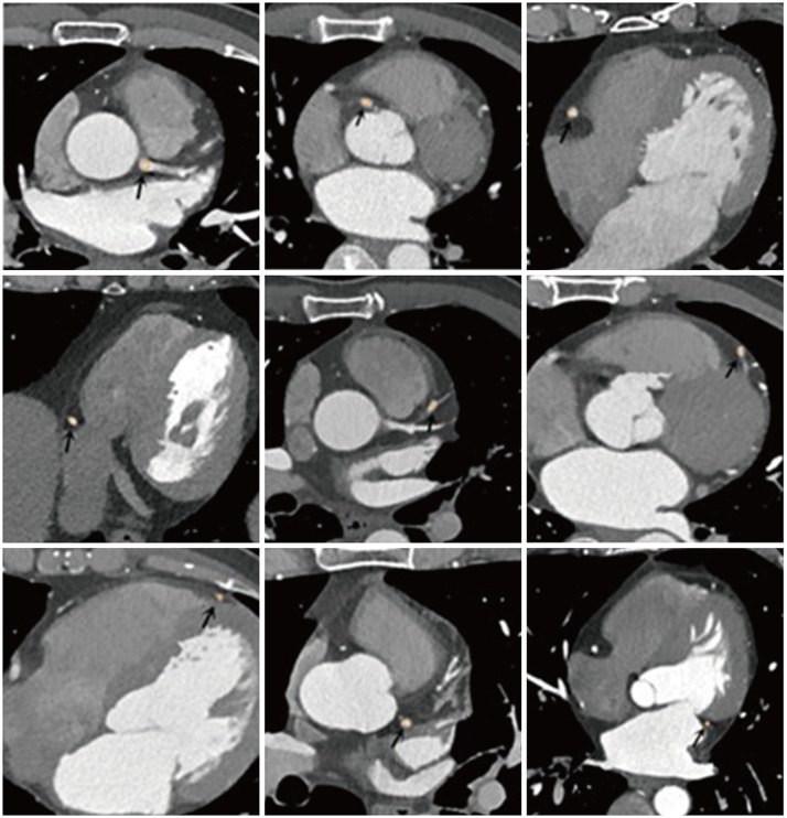
Fig. 1 CT value measurement in coronary artery segments (arrows) on IMR images.CT values were measured in lumen of left main trunk, proximal, middle, and distal segments of right coronary artery, LAD artery, and proximal and distal segments of left circumflex artery. IMR = iterative model reconstruction, LAD = left anterior descending
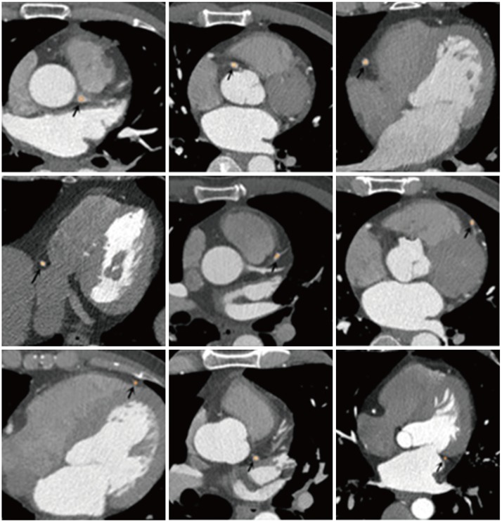
Fig. 2 CT value measurement in coronary artery segments (arrows) on HIR images in same patient.HIR = hybrid iterative reconstruction

Fig. 3 Plaque identification and CT value measurement.A. Non-calcified plaque was detected on proximal segment of LAD on curved reformation. B. CT value of plaque and adjacent perivascular adipose tissue measurement. Ar = area, Av = average, Max = maximum, Min = minimum, SD = standard deviation
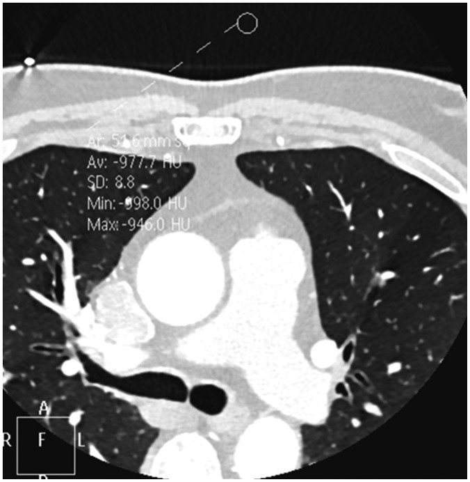
Fig. 4 Noise measurement.Noise was defined as SD measured in background (air) anterior to patients' chest wall. Area of region of interest for measuring noise was about 50 mm2.
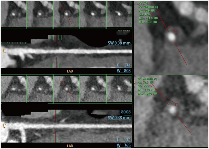
Fig. 5 Representative figures of napkin-ring sign in 47-year-old male patient.Top figures show that napkin-ring sign of LAD plaque can be detected using IMR algorithm, but not on HIR image (bottom figures).
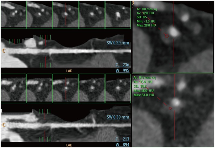
Fig. 6 Representative figures of napkin-ring sign in 64-year-old female patient.Top figures show that napkin-ring sign of LAD plaque can be detected using IMR algorithm, but not on HIR image (bottom figures).
Reference
-
1. Smith-Bindman R. Is computed tomography safe? N Engl J Med. 2010; 363:1–4. PMID: 20573919.
Article2. Leipsic J, Heilbron BG, Hague C. Iterative reconstruction for coronary CT angiography: finding its way. Int J Cardiovasc Imaging. 2012; 28:613–620. PMID: 21359835.
Article3. Silva AC, Lawder HJ, Hara A, Kujak J, Pavlicek W. Innovations in CT dose reduction strategy: application of the adaptive statistical iterative reconstruction algorithm. AJR Am J Roentgenol. 2010; 194:191–199. PMID: 20028923.
Article4. Hou Y, Liu X, Xv S, Guo W, Guo Q. Comparisons of image quality and radiation dose between iterative reconstruction and filtered back projection reconstruction algorithms in 256-MDCT coronary angiography. AJR Am J Roentgenol. 2012; 199:588–594. PMID: 22915398.
Article5. Klink T, Obmann V, Heverhagen J, Stork A, Adam G, Begemann P. Reducing CT radiation dose with iterative reconstruction algorithms: the influence of scan and reconstruction parameters on image quality and CTDIvol. Eur J Radiol. 2014; 83:1645–1654. PMID: 25037931.
Article6. Leipsic J, Labounty TM, Heilbron B, Min JK, Mancini GB, Lin FY, et al. Adaptive statistical iterative reconstruction: assessment of image noise and image quality in coronary CT angiography. AJR Am J Roentgenol. 2010; 195:649–654. PMID: 20729442.
Article7. Kröpil P, Bigdeli AH, Nagel HD, Antoch G, Cohnen M. Impact of increasing levels of advanced iterative reconstruction on image quality in low-dose cardiac CT angiography. Rofo. 2014; 186:567–575. PMID: 24458375.8. Wang R, Schoepf UJ, Wu R, Reddy RP, Zhang C, Yu W, et al. Image quality and radiation dose of low dose coronary CT angiography in obese patients: sinogram affirmed iterative reconstruction versus filtered back projection. Eur J Radiol. 2012; 81:3141–3145. PMID: 22578834.
Article9. Yuki H, Utsunomiya D, Funama Y, Tokuyasu S, Namimoto T, Hirai T, et al. Value of knowledge-based iterative model reconstruction in low-kV 256-slice coronary CT angiography. J Cardiovasc Comput Tomogr. 2014; 8:115–123. PMID: 24661824.
Article10. Halpern EJ, Gingold EL, White H, Read K. Evaluation of coronary artery image quality with knowledge-based iterative model reconstruction. Acad Radiol. 2014; 21:805–811. PMID: 24809321.
Article11. Oda S, Weissman G, Vembar M, Weigold WG. Iterative model reconstruction: improved image quality of low-tube-voltage prospective ECG-gated coronary CT angiography images at 256-slice CT. Eur J Radiol. 2014; 83:1408–1415. PMID: 24873832.
Article12. Oda S, Utsunomiya D, Funama Y, Katahira K, Honda K, Tokuyasu S, et al. A knowledge-based iterative model reconstruction algorithm: can super-low-dose cardiac CT be applicable in clinical settings? Acad Radiol. 2014; 21:104–110. PMID: 24331272.13. Zhang F, Yang L, Song X, Li YN, Jiang Y, Zhang XH, et al. Feasibility study of low tube voltage (80 kVp) coronary CT angiography combined with contrast medium reduction using iterative model reconstruction (IMR) on standard BMI patients. Br J Radiol. 2016; 89:20150766. PMID: 26607646.14. Lee J, Park CH, Oh CS, Han K, Kim TH. Coronary computed tomographic angiography at 80 kVp and knowledge-based iterative model reconstruction is non-inferior to that at 100 kVp with iterative reconstruction. PLoS One. 2016; 11:e0163410. PMID: 27658197.
Article15. Virmani R, Burke AP, Farb A, Kolodgie FD. Pathology of the vulnerable plaque. J Am Coll Cardiol. 2006; 47(8 Suppl):C13–C18. PMID: 16631505.
Article16. Leipsic J, Abbara S, Achenbach S, Cury R, Earls JP, Mancini GJ, et al. SCCT guidelines for the interpretation and reporting of coronary CT angiography: a report of the Society of Cardiovascular Computed Tomography Guidelines Committee. J Cardiovasc Comput Tomogr. 2014; 8:342–358. PMID: 25301040.
Article17. Feuchtner G, Kerber J, Burghard P, Dichtl W, Friedrich G, Bonaros N, et al. The high-risk criteria low-attenuation plaque <60 HU and the napkin-ring sign are the most powerful predictors of MACE: a long-term follow-up study. Eur Heart J Cardiovasc Imaging. 2017; 18:772–779. PMID: 27502292.18. Schmid M, Pflederer T, Jang IK, Ropers D, Sei K, Daniel WG, et al. Relationship between degree of remodeling and CT attenuation of plaque in coronary atherosclerotic lesions: an in-vivo analysis by multi-detector computed tomography. Atherosclerosis. 2008; 197:457–464. PMID: 17727859.
Article19. Hu MQ, Li M, Liu ZY, Huang MP, Liu H, Liang CH. Image quality evaluation of iterative model reconstruction on low tube voltage (80 kVp) coronary CT angiography in an animal study. Acta Radiol. 2016; 57:170–177. PMID: 25657261.
Article20. Chang W, Lee JM, Lee K, Yoon JH, Yu MH, Han JK, et al. Assessment of a model-based, iterative reconstruction algorithm (MBIR) regarding image quality and dose reduction in liver computed tomography. Invest Radiol. 2013; 48:598–606. PMID: 23511193.
Article21. Hausleiter J, Martinoff S, Hadamitzky M, Martuscelli E, Pschierer I, Feuchtner GM, et al. Image quality and radiation exposure with a low tube voltage protocol for coronary CT angiography results of the PROTECTION II Trial. JACC Cardiovasc Imaging. 2010; 3:1113–1123. PMID: 21070998.22. Feuchtner GM, Jodocy D, Klauser A, Haberfellner B, Aglan I, Spoeck A, et al. Radiation dose reduction by using 100-kV tube voltage in cardiac 64-slice computed tomography: a comparative study. Eur J Radiol. 2010; 75:e51–e56. PMID: 19671491.
Article23. Park CH, Lee J, Oh C, Han KH, Kim TH. The feasibility of sub-millisievert coronary CT angiography with low tube voltage, prospective ECG gating, and a knowledge-based iterative model reconstruction algorithm. Int J Cardiovasc Imaging. 2015; 31(Suppl 2):197–203. PMID: 26521066.
Article24. André F, Fortner P, Vembar M, Mueller D, Stiller W, Buss SJ, et al. Improved image quality with simultaneously reduced radiation exposure: knowledge-based iterative model reconstruction algorithms for coronary CT angiography in a clinical setting. J Cardiovasc Comput Tomogr. 2017; 11:213–220. PMID: 28314613.
Article25. Iyama Y, Nakaura T, Yokoyama K, Kidoh M, Harada K, Oda S, et al. Low-contrast and low-radiation dose protocol in cardiac computed tomography: usefulness of low tube voltage and knowledge-based iterative model reconstruction algorithm. J Comput Assist Tomogr. 2016; 40:941–947. PMID: 27224224.
- Full Text Links
-
- Actions
-
Cited
- CITED
-
- Close
- Share
-
- Similar articles
-
- Calcified Plaque of Coronary Artery: Factors Influencing Overestimation of Coronary Artery Stenosis on Coronary CT Angiography
- Noninvasive Detection of Coronary Atherosclerotic Plaques and Assessment of Stenosis Degree at Multidetector CT Coronary Angiography
- Coronary Stent on Coronary CT Angiography: Assessment with Model-Based Iterative Reconstruction Technique
- Plaque Characteristics on Coronary CT Angiography: Effects on the Degree of Coronary Artery Stenosis
- Erratum: Coronary Stent on Coronary CT Angiography: Assessment with Model-Based Iterative Reconstruction Technique
