A Case of Perimembranous Ventricular Septal Defect Associated with Sinus of Valsalva Aneurysm Mimicking Membranous Septal Aneurysm
- Affiliations
-
- 1Department of Internal Medicine, Ulsan University Hospital, University of Ulsan College of Medicine, Ulsan, Korea. kimsc226@gmail.com
- 2Department of Cardiac Surgery, Ulsan University Hospital, University of Ulsan College of Medicine, Ulsan, Korea.
- KMID: 1806922
- DOI: http://doi.org/10.4250/jcu.2015.23.2.113
Abstract
- Sinus of Valsalva aneurysms are rare. Sinus of Valsalva aneurysms are frequently associated with ventricular septal defect (VSD) and aortic regurgitation. They often remain asymptomatic until abruptly presenting with acute chest pain and heart failure secondary to rupture. Here, we describe a case of 20-year-old man who presented with chest pain with a history of VSD. Initial work-up concluded that the patient had VSD associated membranous septal aneurysm. Four years later, the patient presented with symptoms of heart failure. Work-up showed that the ruptured sinus of Valsalva aneurysm was the cause of symptoms. Due to its close proximity to the aortic annulus, sinus of Valsalva aneurysm should be differentiated from membranous septal aneurysm.
Keyword
MeSH Terms
Figure
-
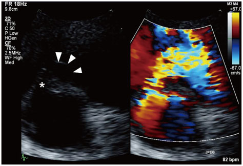
Fig. 1 Initial transthoracic echocardiography. At parasternal short axis view, perimembranous ventricular septal defect (star) was detected with 2 dimensional and color Doppler echocardiography. Focal aneurysmal dilatation (arrowheads) was noted around the defect.
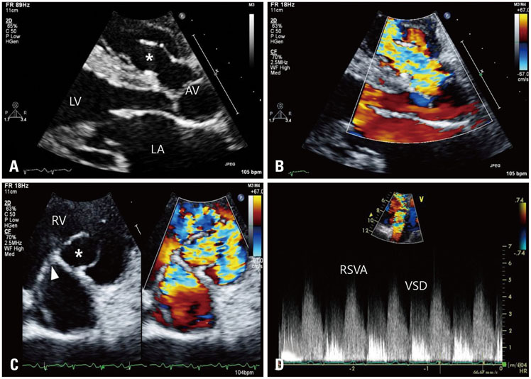
Fig. 2 Transthoracic echocardiography performed at the emergency room. A: At parasternal long axis view, 2-dimensional imaging showed the "windsock" appearance dilatation of right sinus of Valsalva (star) and its tip was ruptured. B: Color Doppler imaging showed diastolic jet flow through the ruptured right sinus of Valsalva aneurysm from the aortic root to the right ventricle. C: At parasternal short axis view, 2-dimensional and color Doppler imaging showed the RSVA (star) and VSD (arrowhead). D: At apical 5 chamber view, continuous wave Doppler showed systolic jet from VSD and diastolic jet from RSVA clearly. AV: aortic valve, LA: left atrium, LV: left ventricle, RV: right ventricle, RSVA: ruptured sinus of Valsalva aneurysm, VSD: ventricular septal defect.
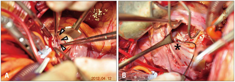
Fig. 3 Photographs of surgical repair. A: After aortotomy, there was small defect at the tip of sinus of Valsalva aneurysm (8 mm sized, arrowheads). B: After right ventriculotomy, probe indicated the perimembranous ventricular septal defect (1 cm sized, star).

Fig. 4 Comparative images between sinus of Valsalva aneurysm and membranous septal aneurysm. A: Current case of sinus of Valsalva aneurysm at apical 5 chamber view showed aneurysmal dilatation (star) above the aortic annulus. B: A case of membranous septal aneurysm at apical 4 chamber view showed aneurysmal dilatation (star) below the aortic annulus. Ao: aorta, LA: left atrium, LV: left ventricle, RA: right atrium.
Reference
-
1. Edwards JE, Burchell HB. The pathological anatomy of deficiencies between the aortic root and the heart, including aortic sinus aneurysms. Thorax. 1957; 12:125–139.
Article2. Michael DF, William HP. A sinus of valsalva fistula. In : Alexander RW, Schlant RC, Fuster V, editors. Hurst's the heart, arteries and veins. 9th ed. New York: McGraw-Hill;1998.3. Lee TH, Lee DW, Cho JY, Hyun MC, Lee SB. Clinical features and surgical results of ruptured sinus of valsalva aneurysm. Korean J Pediatr. 2006; 49:287–291.
Article4. Moon KS, Choi RK, Lim DS, Park HS, Hong SK, Lee YT, Hwang HK. Clinical characteristics in patients with ruptured aneurysm of sinus of Valsalva. Korean Circ J. 2000; 30:183–190.
Article5. De Bakey ME, Diethrich EB, Liddicoat JE, Kinard SA, Garrett HE. Abnormalities of the sinuses of valsalva. Experience with 35 patients. J Thorac Cardiovasc Surg. 1967; 54:312–332.6. Sanchez HE, Barnard CN, Barnard MS. Fistula of the sinus of Valsalva. J Thorac Cardiovasc Surg. 1977; 73:877–879.
Article7. Takach TJ, Reul GJ, Duncan JM, Cooley DA, Livesay JJ, Ott DA, Frazier OH. Sinus of Valsalva aneurysm or fistula: management and outcome. Ann Thorac Surg. 1999; 68:1573–1577.
Article8. Chu SH, Hung CR, How SS, Chang H, Wang SS, Tsai CH, Liau CS, Tseng CD, Tseng YZ, Lee YT, et al. Ruptured aneurysms of the sinus of Valsalva in Oriental patients. J Thorac Cardiovasc Surg. 1990; 99:288–298.
Article9. Norwicki ER, Aberdeen E, Friedman S, Rashkind WJ. Congenital left aortic sinus-left ventricle fistula and review of aortocardiac fistulas. Ann Thorac Surg. 1977; 23:378–388.
Article10. Burakovsky VI, Podsolkov VP, Sabirow BN, Nasedkina MA, Alekian BG, Dvinyaninova NB. Ruptured congenital aneurysm of the sinus of Valsalva. Clinical manifestations, diagnosis, and results of surgical corrections. J Thorac Cardiovasc Surg. 1988; 95:836–841.11. van Son JA, Danielson GK, Schaff HV, Orszulak TA, Edwards WD, Seward JB. Long-term outcome of surgical repair of ruptured sinus of Valsalva aneurysm. Circulation. 1994; 90(5 Pt 2):II20–II29.12. Joo SJ, Koh KG, Kim YH, Park YB, Choi YS, Seo JD, Lee YW, Park JH, Suh KP. Clinical observation on ruptured aneurysm of the sinus of Valsalva. Korean Circ J. 1987; 17:149–158.
Article13. Acar P, Abadir S, Aggoun Y. Transcatheter closure of perimembranous ventricular septal defects with Amplatzer occluder assessed by real-time three-dimensional echocardiography. Eur J Echocardiogr. 2007; 8:110–115.
Article14. Schmaltz AA, Schaefer M, Hentrich F, Neudorf U, Brecher AM, Asfour B, Urban AE. [Ventricular septal defect and aortic regurgitation-pathophysiological aspects and therapeutic consequences]. Z Kardiol. 2004; 93:194–200.15. Eapen RS, Lemler MS, Scott WA, Ramaciotti C. Echocardiographic characteristics of perimembranous ventricular septal defects associated with aortic regurgitation. J Am Soc Echocardiogr. 2003; 16:209–213.
Article16. Dev V, Goswami KC, Shrivastava S, Bahl VK, Saxena A. Echocardiographic diagnosis of aneurysm of the sinus of Valsalva. Am Heart J. 1993; 126:930–936.
Article17. Terdjman M, Bourdarias JP, Farcot JC, Gueret P, Dubourg O, Ferrier A, Hanania G. Aneurysms of sinus of Valsalva: two-dimensional echocardiographic diagnosis and recognition of rupture into the right heart cavities. J Am Coll Cardiol. 1984; 3:1227–1235.
Article18. Wang KY, St John, Ho HY, Ting CT. Congenital sinus of Valsalva aneurysm: a multiplane transesophageal echocardiographic experience. J Am Soc Echocardiogr. 1997; 10:956–963.
Article19. Choi M, Jung JI, Lee BY, Kim HR. Ventricular septal aneurysms in adults: findings of cardiac CT images and correlation with clinical features. Acta Radiol. 2011; 52:619–623.
Article20. Beerman LB, Park SC, Fischer DR, Fricker FJ, Mathews RA, Neches WH, Lenox CC, Zuberbuhler JR. Ventricular septal defect associated with aneurysm of the membranous septum. J Am Coll Cardiol. 1985; 5:118–123.
Article
- Full Text Links
-
- Actions
-
Cited
- CITED
-
- Close
- Share
-
- Similar articles
-
- A Case of Ruptured Aneurysm of Right Sinus of Valsalva Diagnosed Preoperatively by Echocardiography
- Unruptured right sinus of Valsalva aneurysm in a Maltese dog: a case report
- A Rare Case of Unruptured Sinus of Valsalva Aneurysm Obstructing the Right Ventricular Outflow Tract
- Embolization of the Device to the Left Pulmonary Artery after the Interventional Closure of Ruptured Sinus of Valsalva Aneurysm
- Anomaly of the Left Anterior Descending Coronary Artery Arising from the Right Sinus of Valsalva and Ventricular Septal Defect in Adult: A Rare Case
