A Rare Case of Unruptured Sinus of Valsalva Aneurysm Obstructing the Right Ventricular Outflow Tract
- Affiliations
-
- 1Division of Cardiology, Department of Internal Medicine, Presbyterian Medical Center, Jeonju, Korea. john-sh@hanmail.net
- KMID: 2135439
- DOI: http://doi.org/10.4250/jcu.2010.18.2.55
Abstract
- An unruptured sinus of Valsalva aneurysm is rare and is usually asymptomatic until a symptom associated with its complication develops. Hence, an unruptured sinus of Valsalva aneurysm is not infrequently missed unless echocardiogram is performed with other indications. An unruptured sinus of Valsalva aneurysm rarely protrudes into the right ventricular outflow tract, causing the right ventricular outflow tract obstruction. In this report, I describe a rare case of unruptured sinus of Valsalva aneurysm producing the right ventricular outflow tract obstruction, which was incidentally detected by echocardiography.
Figure
-
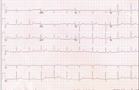
Fig. 1 Electrocardiogram shows normal sinus rhythm.
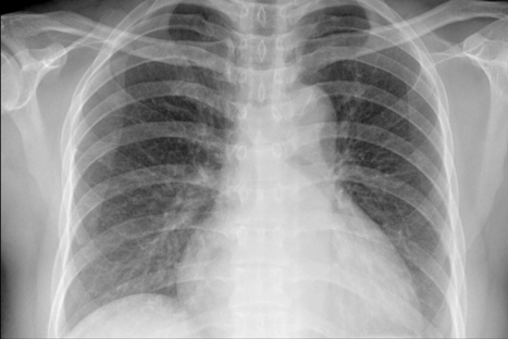
Fig. 2 Chest X-ray shows mild cardiomegaly.

Fig. 3 Transthoracic echocardiography. Parasternal long axis view (A) shows an aneurysm of the right sinus of Valsalva (arrow). Parasternal short axis views (B and C) show also the aneurysm (arrowheads) and systolic flow acceleration suggesting the RVOT obstruction (arrow). RVOT: right ventricular outflow tract, LV: left ventricle, LA: left atrium, Ao: aorta.

Fig. 4 Transesophageal echocardiography. Midesophageal aortic valve long-axis view at 144℃ rotation demonstrating the SVA originating from the right coronary sinus of Valsalva (arrow) (A). Short-axis view at 27℃ rotation demonstrating the SVA protruding into the RVOT (arrowheads) (B). Color Doppler shows flow acceleration due to the RVOT obstruction without shunt flow suggesting SVA rupture (arrow) (C). SVA: sinus of Valsalva aneurysm, RVOT: right ventricular outflow tract, LV: left ventricle, LA: left atrium, Ao: aorta.
Reference
-
1. Goldberg N, Krasnow N. Sinus of Valsalva aneurysms. Clin Cardiol. 1990. 13:831–836.
Article2. Rosenberger P, Cohn LH, Fox JA, Locke A, Shernan SK. Sinus of Valsalva aneurysm obstructing the right ventricular outflow tract. Anesth Analg. 2006. 102:1660–1661.
Article3. Freedom RM, Yoo SJ. Freedom RM, Yoo SJ, Mikailian H, William WG, editors. Sinus of Valsalva aneurysm. The natural and modified history of congenital heart disease. 2004. 1st Edition. New York: Blackwell Publishing;183–185.
Article4. Vereckei A, Vándor L, Halász J, Karádi I, Lengyel M. Infective endocarditis resulting in rupture of sinus of Valsalva with a rupture site communicating with both the right atrium and right ventricle. J Am Soc Echocardiogr. 2004. 17:995–997.
Article5. Bansal RC, Wangsnes KM, Bailey L. Right aortic sinus of Valsalva-to-right ventricle fistula complicating bacterial endocarditis of membranous ventricular septal defect: evaluation by two-dimensional, color flow, and Doppler echocardiography. J Am Soc Echocardiogr. 1993. 6:308–311.
Article6. Chu SH, Hung CR, How SS, Chang H, Wang SS, Tsai CH, Liau CS, Tseng CD, Tseng YZ, Lee YT, et al. Ruptured aneurysms of sinus of Valsalva in Oriental patients. J Thoracic Cardiovasc Surg. 1990. 99:288–298.7. Regueiro Abel M, Penas Lado M, López Ciudad V, Castro Beiras A. [Sinus of Valsalva aneurysm as a cause of acute myocardial infarction.]. Rev Esp Cardiol. 2002. 55:77–79.8. Takach TJ, Reul GJ, Duncan JM, Cooley DA, Livesay JJ, Ott DA, Frazier OH. Sinus of Valsalva aneurysm or fistula: management and outcome. Ann Thorac Surg. 1999. 68:1573–1577.
Article9. Vural KM, Sener E, Taşdemir O, Bayazit K. Approach to sinus of Valsalva aneurysm: a review of 53 cases. Eur J Cardiothorac Surg. 2001. 20:71–76.
- Full Text Links
-
- Actions
-
Cited
- CITED
-
- Close
- Share
-
- Similar articles
-
- A Case of Unruptured Aneurysm of the Right Sinus of Valsalva with Right Ventricular Outflow Obstruction
- A Case of Right Sinus of Valsalva Rupture with Dissection into Interventricular Septum Causing Left Ventricular Outflow Tract Obstruction
- Unruptured right sinus of Valsalva aneurysm in a Maltese dog: a case report
- A Giant Aneurysm of the Sinus of Valsalva with Calcification
- A Giant Unruptured Right Coronary Sinus of Valsalva Aneurysm
