Clinical and Radiological Predictive Factors to be Related with the Degree of Lumbar Back Muscle Degeneration: Difference by Gender
- Affiliations
-
- 1Department of Orthopaedic Surgery, Kangwon National University School of Medicine, Chuncheon, Korea. spinecjh@gmail.com
- 2Department of Orthopedic Surgery, Seoul National University College of Medicine, Seoul, Korea.
- KMID: 1794714
- DOI: http://doi.org/10.4055/cios.2014.6.3.318
Abstract
- BACKGROUND
The prediction of lumbar back muscle degeneration is important because chronic low back pain and spino-pelvic imbalance have been known to be related to it. However, gender difference should be considered because there are different quality and volume of muscles between genders. The purpose of this study was to search for clinical and radiological factors to predict the degree of lumbar back muscle degeneration according to gender difference.
METHODS
We reviewed 112 patients (44 men and 68 women) with spinal stenosis who underwent a decompressive surgery between 1 January 2009 and 31 December 2011. Degrees of lumbar back muscle degeneration were classified into three categories by the fatty infiltration at each L3-4 disc level on the axial view of T1 magnetic resonance imaging (MRI). Age, sex, bone marrow density score, and body mass index (BMI) were obtained from chart reviews. Lumbar lordosis, sacral slope, pelvic tilt (PT), and pelvic incidence were calculated with lumbar spine standing lateral radiographs. The degrees of spinal stenosis and facet arthropathy were checked with MRI. Student t-test, chi-square test, or Fisher exact test were used to compare clinical and radiological parameters between genders. Analysis of variance (ANOVA) and linear regression analysis were used to search for a relationship between lumbar back muscle degeneration and possible predictive factors in each gender group.
RESULTS
Many clinical and radiological parameters were different according to gender. The age, BMI, and PT in the female group (p = 0.013, 0.001, and 0.019, respectively) and the PT in the men group (p = 0.018) were predictive factors to be correlated with lumbar back muscle degeneration.
CONCLUSIONS
The PT was the important predictive factor for lumbar back muscle degeneration in both, the male and the female group. However, age and BMI were predictive factors in the female group only.
MeSH Terms
-
Aged
Back Muscles/*pathology/physiopathology/radiography
Chronic Disease
Decompression, Surgical
Female
Humans
Low Back Pain/*diagnosis/physiopathology/surgery
Lumbosacral Region
Magnetic Resonance Imaging
Male
Middle Aged
Postural Balance
Posture
Predictive Value of Tests
Retrospective Studies
Spinal Stenosis/*diagnosis/physiopathology/surgery
Figure
-
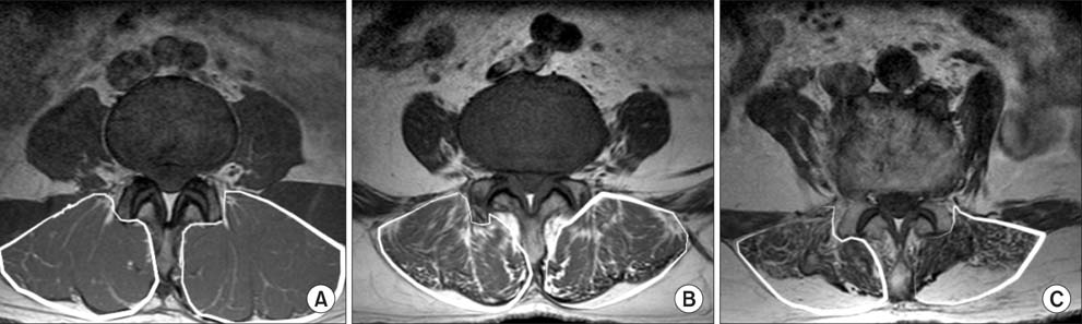
Fig. 1 Qualitative analysis of the fatty infiltration of lumbar back muscles including both multifidus and longissimus (demarcated by white lines) on T1 axial section of magnetic resonance imaging of the L3-4 disc level using a three tier grading system. (A) Grade 1: mild degree (< 10% fatty infiltration). (B) Grade 2: moderate degree (10%-50% fatty infiltration). (C) Grade 3: severe degree (> 50% fatty infiltration).
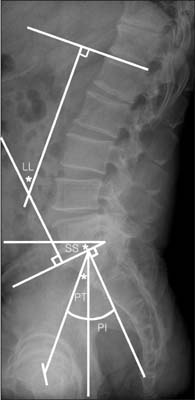
Fig. 2 Illustration of the measurement of spino-pelvic parameters including pelvic tilt (PT), sacral slope (SS), pelvic incidence (PI), and lumbar lordosis (LL) on lumbar standing lateral radiographs.
Cited by 1 articles
-
Effect of Sagittal Balance on Risk of Falling after Lateral Lumbar Interbody Fusion Surgery Combined with Posterior Surgery
Byung Ho Lee, Jae-Ho Yang, Hak-Sun Kim, Kyung-Soo Suk, Hwan-Mo Lee, Jin-Oh Park, Seong-Hwan Moon
Yonsei Med J. 2017;58(6):1177-1185. doi: 10.3349/ymj.2017.58.6.1177.
Reference
-
1. Mengiardi B, Schmid MR, Boos N, et al. Fat content of lumbar paraspinal muscles in patients with chronic low back pain and in asymptomatic volunteers: quantification with MR spectroscopy. Radiology. 2006; 240(3):786–792.2. Kang CH, Shin MJ, Kim SM, Lee SH, Lee CS. MRI of paraspinal muscles in lumbar degenerative kyphosis patients and control patients with chronic low back pain. Clin Radiol. 2007; 62(5):479–486.3. Lee JC, Cha JG, Kim Y, Kim YI, Shin BJ. Quantitative analysis of back muscle degeneration in the patients with the degenerative lumbar flat back using a digital image analysis: comparison with the normal controls. Spine (Phila Pa 1976). 2008; 33(3):318–325.4. Imagama S, Matsuyama Y, Hasegawa Y, et al. Back muscle strength and spinal mobility are predictors of quality of life in middle-aged and elderly males. Eur Spine J. 2011; 20(6):954–961.5. Kawakami M, Tamaki T, Ando M, Yamada H, Hashizume H, Yoshida M. Lumbar sagittal balance influences the clinical outcome after decompression and posterolateral spinal fusion for degenerative lumbar spondylolisthesis. Spine (Phila Pa 1976). 2002; 27(1):59–64.6. Goutallier D, Postel JM, Bernageau J, Lavau L, Voisin MC. Fatty muscle degeneration in cuff ruptures: pre- and postoperative evaluation by CT scan. Clin Orthop Relat Res. 1994; (304):78–83.7. Doherty TJ. The influence of aging and sex on skeletal muscle mass and strength. Curr Opin Clin Nutr Metab Care. 2001; 4(6):503–508.8. Mannion AF, Dumas GA, Cooper RG, Espinosa FJ, Faris MW, Stevenson JM. Muscle fibre size and type distribution in thoracic and lumbar regions of erector spinae in healthy subjects without low back pain: normal values and sex differences. J Anat. 1997; 190(Pt 4):505–513.9. Kjaer P, Bendix T, Sorensen JS, Korsholm L, Leboeuf-Yde C. Are MRI-defined fat infiltrations in the multifidus muscles associated with low back pain? BMC Med. 2007; 5:2.10. Schizas C, Theumann N, Burn A, et al. Qualitative grading of severity of lumbar spinal stenosis based on the morphology of the dural sac on magnetic resonance images. Spine (Phila Pa 1976). 2010; 35(21):1919–1924.11. Fujiwara A, Tamai K, Yamato M, et al. The relationship between facet joint osteoarthritis and disc degeneration of the lumbar spine: an MRI study. Eur Spine J. 1999; 8(5):396–401.12. Hultman G, Nordin M, Saraste H, Ohlsen H. Body composition, endurance, strength, cross-sectional area, and density of MM erector spinae in men with and without low back pain. J Spinal Disord. 1993; 6(2):114–123.13. Hides JA, Stokes MJ, Saide M, Jull GA, Cooper DH. Evidence of lumbar multifidus muscle wasting ipsilateral to symptoms in patients with acute/subacute low back pain. Spine (Phila Pa 1976). 1994; 19(2):165–172.14. Barker KL, Shamley DR, Jackson D. Changes in the cross-sectional area of multifidus and psoas in patients with unilateral back pain: the relationship to pain and disability. Spine (Phila Pa 1976). 2004; 29(22):E515–E519.15. Motosuneya T, Asazuma T, Tsuji T, Watanabe H, Nakayama Y, Nemoto K. Postoperative change of the cross-sectional area of back musculature after 5 surgical procedures as assessed by magnetic resonance imaging. J Spinal Disord Tech. 2006; 19(5):318–322.16. Keller A, Gunderson R, Reikeras O, Brox JI. Reliability of computed tomography measurements of paraspinal muscle cross-sectional area and density in patients with chronic low back pain. Spine (Phila Pa 1976). 2003; 28(13):1455–1460.17. D'hooge R, Cagnie B, Crombez G, Vanderstraeten G, Dolphens M, Danneels L. Increased intramuscular fatty infiltration without differences in lumbar muscle cross-sectional area during remission of unilateral recurrent low back pain. Man Ther. 2012; 17(6):584–588.18. Kalichman L, Hodges P, Li L, Guermazi A, Hunter DJ. Changes in paraspinal muscles and their association with low back pain and spinal degeneration: CT study. Eur Spine J. 2010; 19(7):1136–1144.19. Kamaz M, Kiresi D, Oguz H, Emlik D, Levendoglu F. CT measurement of trunk muscle areas in patients with chronic low back pain. Diagn Interv Radiol. 2007; 13(3):144–148.20. Mirtz TA, Greene L. Is obesity a risk factor for low back pain? An example of using the evidence to answer a clinical question. Chiropr Osteopat. 2005; 13(1):2.21. Leboeuf-Yde C. Body weight and low back pain: a systematic literature review of 56 journal articles reporting on 65 epidemiologic studies. Spine (Phila Pa 1976). 2000; 25(2):226–237.22. Lee CS, Lee CK, Kim YT, Hong YM, Yoo JH. Dynamic sagittal imbalance of the spine in degenerative flat back: significance of pelvic tilt in surgical treatment. Spine (Phila Pa 1976). 2001; 26(18):2029–2035.23. Gottfried ON, Daubs MD, Patel AA, Dailey AT, Brodke DS. Spinopelvic parameters in postfusion flatback deformity patients. Spine J. 2009; 9(8):639–647.24. Kim H, Lee CK, Yeom JS, et al. Asymmetry of the cross-sectional area of paravertebral and psoas muscle in patients with degenerative scoliosis. Eur Spine J. 2013; 22(6):1332–1338.
- Full Text Links
-
- Actions
-
Cited
- CITED
-
- Close
- Share
-
- Similar articles
-
- Relationship between Lumbar Disc Degeneration and Back Muscle Degeneration
- Sarcopenia and Back Muscle Degeneration as Risk Factors for Back Pain: A Comparative Study
- Scoring System for Factors Affecting Aggravation of Lumbar Disc Herniation
- Clinical and Radiologic Factors Predicting the Low Back Pain after Discectomy
- Atrophy of Multifidus Muscle on Low Back Pain Patients
