Scoring System for Factors Affecting Aggravation of Lumbar Disc Herniation
- Affiliations
-
- 1Department of Radiology, Dankook University Hospital, Cheonan, Korea. mirrorartifact@dkuh.co.kr
- KMID: 2408814
- DOI: http://doi.org/10.13104/imri.2018.22.1.18
Abstract
- PURPOSE
To investigate the various imaging factors associated with aggravation of lumbar disc herniation (LDH) and develop a scoring system for prediction of LDH aggravation.
MATERIALS AND METHODS
From 2015 to 2017, we retrospectively reviewed the magnetic resonance imaging (MRI) findings of 60 patients (30 patients with aggravated LDH and 30 patients without any altered LDH). Imaging factors for MRI evaluation included the level of LDH, disc degeneration, back muscle atrophy, facet joint degeneration, ligamentum flavum thickness and interspinous ligament degeneration. Flexion-extension difference was measured with simple radiography. The scoring system was analyzed using receiver operating characteristic (ROC) analysis.
RESULTS
The aggravated group manifested a higher grade of disc degeneration, back muscle atrophy and facet degeneration than the control group. The ligamentum flavum thickness in the aggravated group was thicker than in the group with unaltered LDH. The summation score was defined as the sum of the grade of disc degeneration, back muscle atrophy and facet joint degeneration. The area under the ROC curve showing the threshold value of the summation score for prediction of aggravation of LDH was 0.832 and the threshold value corresponded to 6.5.
CONCLUSION
Disc degeneration, facet degeneration, back muscle atrophy and ligamentum flavum thickness are important factors in predicting aggravation of LDH and may facilitate the determination of treatment strategy in patients with LDH. The summation score is available as supplemental data.
Keyword
MeSH Terms
Figure
-

Fig. 1. Measurement perpendicular to the thickest portion of ligamentum flavum at the herniated disc level on T2-weighted image axial scan.

Fig. 2. The flexion-extension difference was measured on lateral simple radiograph.

Fig. 3. The graph showing the number of patients for each summation score of the Pfirrmann disc degeneration grade, back muscle atrophy, and facet joint degeneration.

Fig. 4. Receiver operating characteristic curve showing the threshold value of the summation score of the Pfirrmann disc degeneration grade, back muscle atrophy, and facet joint degeneration to predict the aggravation of lumbar disc herniation.
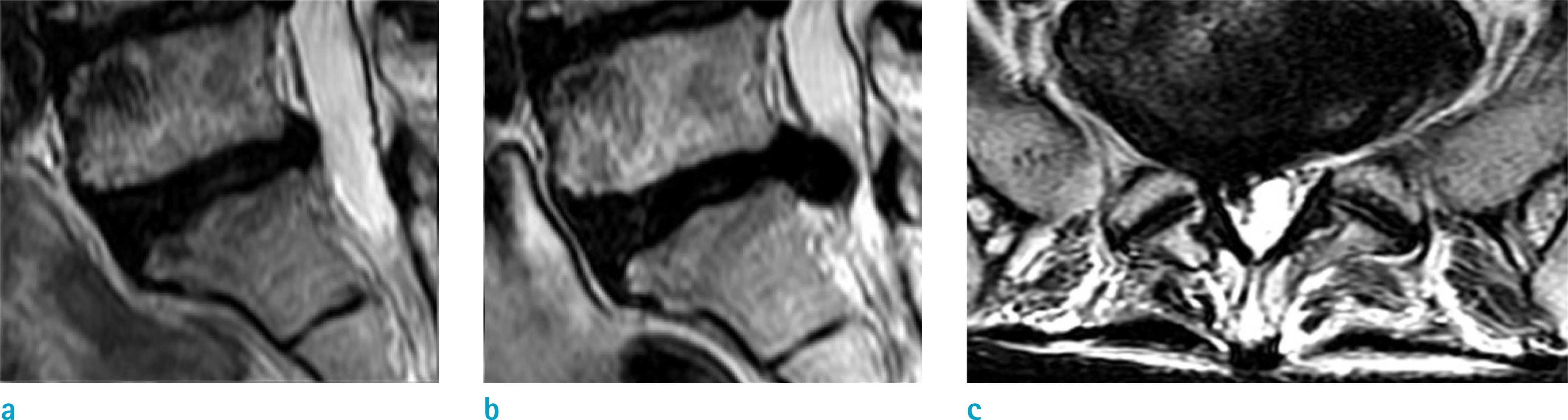
Fig. 5. A 63-year-old woman with right subarticular disc extrusion, L5-S1. (a) Initial sagittal T2-weighted image shows disc degeneration grade 4. (b) Sagittal T2-weighted image shows lumbar disc herniation aggravation after 58 months. (c) Initial axial T2-weighted image shows back muscle atrophy grade 3, facet joint degeneration grade 1 and ligamentum flavum thickness 2.2 mm. The summation score is 8.
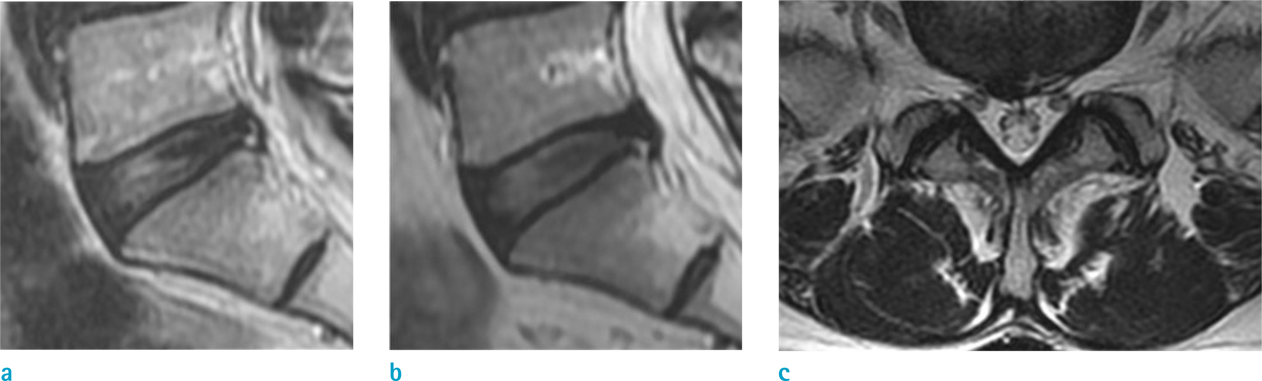
Fig. 6. A 58-year-old woman with right central disc protrusion, L5-S1. (a) Initial sagittal T2-weighted image shows disc degeneration grade 2. (b) Sagittal T2-weighted image shows no significant change in lumbar disc herniation after 47 months. (c) Initial axial T2-weighted image shows grade 1 back muscle atrophy, facet joint degeneration grade 1 and ligamentum flavum thickness 1.9 mm. The summation score is 4.
Reference
-
References
1. Frymoyer JW. Lumbar disk disease: epidemiology. Instr Course Lect. 1992; 41:217–223.2. Fager CA. Observations on spontaneous recovery from intervertebral disc herniation. Surg Neurol. 1994; 42:282286.
Article3. Weber H. Lumbar disc herniation. A controlled, prospective study with ten years of observation. Spine (Phila Pa 1976). 1983; 8:131–140.
Article4. Weber H, Holme I, Amlie E. The natural course of acute sciatica with nerve root symptoms in a double-blind placebo-controlled trial evaluating the effect of piroxicam. Spine (Phila Pa 1976). 1993; 18:1433–1438.
Article5. Hofstee DJ, Gijtenbeek JM, Hoogland PH, et al. Westeinde sciatica trial: randomized controlled study of bed rest and physiotherapy for acute sciatica. J Neurosurg. 2002; 96:45–49.
Article6. Peul WC, van den Hout WB, Brand R, Thomeer RT, Koes BW. Leiden-The Hague Spine Intervention Prognostic Study Group. Prolonged conservative care versus early surgery in patients with sciatica caused by lumbar disc herniation: two year results of a randomised controlled trial. BMJ. 2008; 336:1355–1358.
Article7. Fardon DF, Williams AL, Dohring EJ, Murtagh FR, Gabriel rothman SL, Sze GK. Lumbar disc nomenclature: version 2.0: recommendations of the combined task forces of the North American Spine Society, the American Society of Spine Radiology and the American Society of Neuroradiology. Spine J. 2014; 14:2525–2545.8. Choi SJ, Song JS, Kim C, et al. The use of magnetic resonance imaging to predict the clinical outcome of nonsurgical treatment for lumbar intervertebral disc herniation. Korean J Radiol. 2007; 8:156–163.9. Pfirrmann CW, Metzdorf A, Zanetti M, Hodler J, Boos N. Magnetic resonance classification of lumbar intervertebral disc degeneration. Spine (Phila Pa 1976). 2001; 26:1873–1878.
Article10. Pathria M, Sartoris DJ, Resnick D. Osteoarthritis of the facet joints: accuracy of oblique radiographic assessment. Radiology. 1987; 164:227–230.
Article11. Keorochana G, Taghavi CE, Tzeng ST, et al. MRI classification of interspinous ligament degeneration of the lumbar spine: intraobserver and interobserver reliability and the frequency of disagreement. Eur Spine J. 2010; 19:1740–1745.
Article12. Greiner M, Pfeiffer D, Smith RD. Principles and practical application of the receiver-operating characteristic analysis for diagnostic tests. Prev Vet Med. 2000; 45:23–41.
Article13. Haldeman S. North American Spine Society: failure of the pathology model to predict back pain. Spine (Phila Pa 1976). 1990; 15:718–724.14. Boden SD, Davis DO, Dina TS, Patronas NJ, Wiesel SW. Abnormal magnetic-resonance scans of the lumbar spine in asymptomatic subjects. A prospective investigation. J Bone Joint Surg Am. 1990; 72:403–408.
Article
- Full Text Links
-
- Actions
-
Cited
- CITED
-
- Close
- Share
-
- Similar articles
-
- Clinical Analysis of Recurrent Lumbar Disc Herniation
- Rapid Repeated Recurrent Lumbar Disc Herniation after Microscopic Discectomies
- Juvenile Lumbar Intervertebral Disc Herniation: Five Cases Report
- Redundant Nerve Roots of Cauda Equina Mimicking Intradural Disc Herniation: A Case Report
- High Lumbar Disc Herniation Treated With an Anterolateral Approach: Case Report
