Patient Radiation Dose in Diagnostic and Interventional Procedures for Intracranial Aneurysms: Experience at a Single Center
- Affiliations
-
- 1Department of Radiology, Seoul St. Mary's Hospital, College of Medicine, The Catholic University of Korea, Seoul 137-701, Korea. bumrad@catholic.ac.kr
- 2Department of Neurosurgery, Seoul St. Mary's Hospital, College of Medicine, The Catholic University of Korea, Seoul 137-701, Korea.
- KMID: 1794658
- DOI: http://doi.org/10.3348/kjr.2014.15.6.844
Abstract
OBJECTIVE
To assess patient radiation doses during cerebral angiography and embolization of intracranial aneurysms in a large sample size from a single center.
MATERIALS AND METHODS
We studied a sample of 439 diagnostic and 149 therapeutic procedures for intracranial aneurysms in 480 patients (331 females, 149 males; median age, 57 years; range, 21-88 years), which were performed in 2012 with a biplane unit. Parameters including fluoroscopic time, dose-area product (DAP), and total angiographic image frames were obtained and analyzed.
RESULTS
Mean fluoroscopic time, total mean DAP, and total image frames were 12.6 minutes, 136.6 +/- 44.8 Gy-cm2, and 251 +/- 49 frames for diagnostic procedures, 52.9 minutes, 226.0 +/- 129.2 Gy-cm2, and 241 frames for therapeutic procedures, and 52.2 minutes, 334.5 +/- 184.6 Gy-cm2, and 408 frames for when both procedures were performed during the same session. The third quartiles for diagnostic reference levels (DRLs) were 14.0, 61.1, and 66.1 minutes for fluoroscopy time, 154.2, 272.8, and 393.8 Gy-cm2 for DAP, and 272, 276, and 535 for numbers of image frames in diagnostic, therapeutic, and both procedures in the same session, respectively. The proportions of fluoroscopy in DAP for the procedures were 11.4%, 50.5%, and 36.1%, respectively, for the three groups. The mean DAP for each 3-dimensional rotational angiographic acquisition was 19.2 +/- 3.2 Gy-cm2. On average, rotational angiography was used 1.4 +/- 0.6 times/session (range, 1-4; n = 580).
CONCLUSION
Radiation dose in our study as measured by DAP, fluoroscopy time and image frames did not differ significantly from other reported DRL studies for cerebral angiography, and DAP was lower with fewer angiographic image frames for embolization. A national registry of radiation-dose data is a necessary next step to refine the dose reference level.
Keyword
MeSH Terms
Figure
-
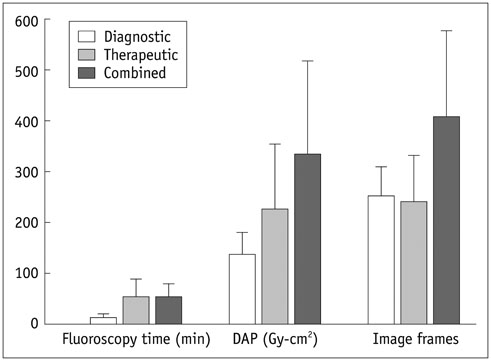
Fig. 1 Chart showing mean of fluoroscopy time, total dose-area product (DAP), and number of image frames among three groups with standard deviation.
Cited by 3 articles
-
Radiation Dose Reduction without Compromise to Image Quality by Alterations of Filtration and Focal Spot Size in Cerebral Angiography
Dong Joon Kim, Min Keun Park, Da Eun Jung, Jung Han Kang, Byung Moon Kim
Korean J Radiol. 2017;18(4):722-728. doi: 10.3348/kjr.2017.18.4.722.Monitoring Radiation Doses during Diagnostic and Therapeutic Neurointerventional Procedures: Multicenter Study for Establishment of Reference Levels
Yon-Kwon Ihn, Bum-soo Kim, Hae Woong Jeong, Sang Hyun Suh, Yoo Dong Won, Young-Jun Lee, Dong Joon Kim, Pyong Jeon, Chang-Woo Ryu, Sang-il Suh, Dae Seob Choi, See Sung Choi, Sang Heum Kim, Jun Soo Byun, Jieun Rho, Yunsun Song, Woo Sang Jeong, Noah Hong, Sung Hyun Baik, Jeong Jin Park, Soo Mee Lim, Jung-Jae Kim, Woong Yoon
Neurointervention. 2021;16(3):240-251. doi: 10.5469/neuroint.2021.00437.Patient Radiation Exposure During Diagnostic and Therapeutic Procedures for Intracranial Aneurysms: A Multicenter Study
Yon Kwon Ihn, Bum-Soo Kim, Jun Soo Byun, Sang Hyun Suh, Yoo Dong Won, Deok Hee Lee, Byung Moon Kim, Young Soo Kim, Pyong Jeon, Chang-Woo Ryu, Sang-il Suh, Dae Seob Choi, See Sung Choi, Jin Wook Choi, Hyuk Won Chang, Jae-Wook Lee, Sang Heum Kim, Young Jun Lee, Shang Hun Shin, Soo Mee Lim, Woong Yoon, Hae Woong Jeong, Moon Hee Han
Neurointervention. 2016;11(2):78-85. doi: 10.5469/neuroint.2016.11.2.78.
Reference
-
1. Bor D, Cekirge S, Türkay T, Turan O, Gülay M, Onal E, et al. Patient and staff doses in interventional neuroradiology. Radiat Prot Dosimetry. 2005; 117:62–68.2. McParland BJ. A study of patient radiation doses in interventional radiological procedures. Br J Radiol. 1998; 71:175–185.3. Struelens L, Vanhavere F, Bosmans H, Van Loon R, Mol H. Skin dose measurements on patients for diagnostic and interventional neuroradiology: a multicentre study. Radiat Prot Dosimetry. 2005; 114:143–146.4. Mahesh M. Fluoroscopy: patient radiation exposure issues. Radiographics. 2001; 21:1033–1045.5. Suzuki S, Furui S, Matsumaru Y, Nobuyuki S, Ebara M, Abe T, et al. Patient skin dose during neuroembolization by multiple-point measurement using a radiosensitive indicator. AJNR Am J Neuroradiol. 2008; 29:1076–1081.6. Aroua A, Rickli H, Stauffer JC, Schnyder P, Trueb PR, Valley JF, et al. How to set up and apply reference levels in fluoroscopy at a national level. Eur Radiol. 2007; 17:1621–1633.7. D'Ercole L, Thyrion FZ, Bocchiola M, Mantovani L, Klersy C. Proposed local diagnostic reference levels in angiography and interventional neuroradiology and a preliminary analysis according to the complexity of the procedures. Phys Med. 2012; 28:61–70.8. Bogaert E, Bacher K, Lemmens K, Carlier M, Desmet W, De Wagter X, et al. A large-scale multicentre study of patient skin doses in interventional cardiology: dose-area product action levels and dose reference levels. Br J Radiol. 2009; 82:303–312.9. Nickoloff EL, Lu ZF, Dutta AK, So JC. Radiation dose descriptors: BERT, COD, DAP, and other strange creatures. Radiographics. 2008; 28:1439–1450.10. Bor D, Sancak T, Olgar T, Elcim Y, Adanali A, Sanlidilek U, et al. Comparison of effective doses obtained from dose-area product and air kerma measurements in interventional radiology. Br J Radiol. 2004; 77:315–322.11. Brambilla M, Marano G, Dominietto M, Cotroneo AR, Carriero A. Patient radiation doses and references levels in interventional radiology. Radiol Med. 2004; 107:408–418.12. Miller DL, Balter S, Cole PE, Lu HT, Schueler BA, Geisinger M, et al. Radiation doses in interventional radiology procedures: the RAD-IR study: part I: overall measures of dose. J Vasc Interv Radiol. 2003; 14:711–727.13. Verdun FR, Aroua A, Trueb PR, Vock P, Valley JF. Diagnostic and interventional radiology: a strategy to introduce reference dose level taking into account the national practice. Radiat Prot Dosimetry. 2005; 114:188–191.14. Le Heron JC. Estimation of effective dose to the patient during medical x-ray examinations from measurements of the dose-area product. Phys Med Biol. 1992; 37:2117–2126.15. Harrison JD, Streffer C. The ICRP protection quantities, equivalent and effective dose: their basis and application. Radiat Prot Dosimetry. 2007; 127:12–18.16. Anxionnat R, Bracard S, Ducrocq X, Trousset Y, Launay L, Kerrien E, et al. Intracranial aneurysms: clinical value of 3D digital subtraction angiography in the therapeutic decision and endovascular treatment. Radiology. 2001; 218:799–808.17. Doelken M, Struffert T, Richter G, Engelhorn T, Nimsky C, Ganslandt O, et al. Flat-panel detector volumetric CT for visualization of subarachnoid hemorrhage and ventricles: preliminary results compared to conventional CT. Neuroradiology. 2008; 50:517–523.18. Kang HS, Han MH, Kwon BJ, Jung SI, Oh CW, Han DH, et al. Postoperative 3D angiography in intracranial aneurysms. AJNR Am J Neuroradiol. 2004; 25:1463–1469.19. Richter G, Engelhorn T, Struffert T, Doelken M, Ganslandt O, Hornegger J, et al. Flat panel detector angiographic CT for stent-assisted coil embolization of broad-based cerebral aneurysms. AJNR Am J Neuroradiol. 2007; 28:1902–1908.20. van Rooij WJ, Sprengers ME, de Gast AN, Peluso JP, Sluzewski M. 3D rotational angiography: the new gold standard in the detection of additional intracranial aneurysms. AJNR Am J Neuroradiol. 2008; 29:976–979.21. Marshall NW, Chapple CL, Kotre CJ. Diagnostic reference levels in interventional radiology. Phys Med Biol. 2000; 45:3833–3846.22. Dekker LR, van der Voort PH, Simmers TA, Verbeek XA, Bullens RW, Veer MV, et al. New image processing and noise reduction technology allows reduction of radiation exposure in complex electrophysiologic interventions while maintaining optimal image quality: a randomized clinical trial. Heart Rhythm. 2013; 10:1678–1682.
- Full Text Links
-
- Actions
-
Cited
- CITED
-
- Close
- Share
-
- Similar articles
-
- Radiation exposure and its reduction in the fluoroscopic examination and fluoroscopy-guided interventional radiology
- Stent Application for the Treatment of Cerebral Aneurysms
- Review of National Diagnostic Reference Levels for Interventional Procedures
- Diagnosis and Treatment of Intracranial Aneurysms with 3-dimensional Digital Subtraction Angiography
- Outpatient Day-care Neuroangiography and Neurointervention of Unruptured Intracranial Aneurysms
