A Case of Residual Non-Small Cell Lung Cancer Cells Coexisting With Newly Developed Small Cell Lung Cancer Cells in Ascitic Fluid after Chemotherapy for Non-Small Cell Lung Cancer
- Affiliations
-
- 1Department of Laboratory Medicine, Pusan National University School of Medicine, Pusan National University Hospital, Busan, Korea.
- 2Department of Laboratory Medicine, University of Ulsan College of Medicine and Asan Medical Center, Seoul, Korea. hschi@amc.seoul.kr
- 3Department of Laboratory Medicine, Kyung Hee University School of Medicine, Seoul, Korea.
- 4Department of Oncology, University of Ulsan College of Medicine and Asan Medical Center, Seoul, Korea.
- KMID: 1791913
- DOI: http://doi.org/10.3343/alm.2014.34.2.163
Abstract
- No abstract available.
MeSH Terms
-
Antineoplastic Agents/therapeutic use
Ascitic Fluid/*cytology
Carcinoma, Non-Small-Cell Lung/*diagnosis/drug therapy/pathology
Humans
Lung Neoplasms/*diagnosis/drug therapy/pathology
Lymphatic Metastasis
Male
Middle Aged
Mutation
Neoplasm Staging
Quinazolines/therapeutic use
Receptor, Epidermal Growth Factor/genetics
Small Cell Lung Carcinoma/*diagnosis/pathology
Tomography, X-Ray Computed
Antineoplastic Agents
Receptor, Epidermal Growth Factor
Quinazolines
Figure
-
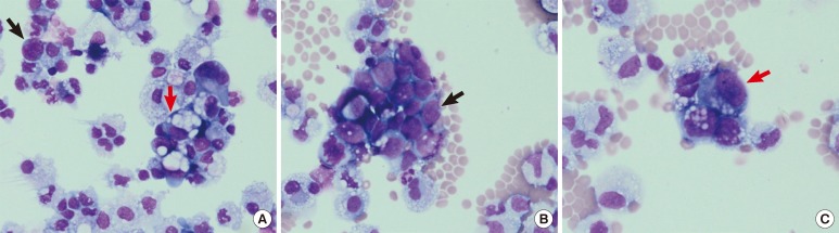
Fig. 1 Wright staining of ascitic fluid cytospin slides. Two types of neoplastic cells were detected (A, 400×, small-cell lung cancer [SCLC] cells pointed by black arrow and non-small cell lung cancer [NSCLC] cells by red arrow) at a frequency of 18%. Small-to medium-sized SCLC cells appeared as clusters of tumor cells with a back-to-back appearance (B, 400×, black arrow), and the second type of cells, large NSCLC cells, showed mucinous contents in the cytoplasm (C, 400×, red arrow).
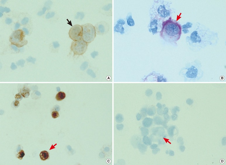
Fig. 2 Cytochemical and immunocytochemical staining results of the two types of neoplastic cells detected in the ascitic fluid. The small-to medium-sized small-cell lung cancer cells (black arrow) showed membrane positivity for CD56 on immunocytochemical staining (A, 400×). The large non-small cell lung cancer cells (red arrow) showed cytoplasmic positivity with periodic acid-Schiff cytochemical staining (B, 1,000×) as well as cytokeratin 7 positivity on immunocytochemical staining (C, 400×) but cytokeratin 20 negativity on immunocytochemical staining (D, 400×).
Reference
-
1. Hiraki A, Ueoka H, Yoshino T, Chikamori K, Onishi K, Kiura K, et al. Synchronous primary lung cancer presenting with small cell carcinoma and non-small cell carcinoma: diagnosis and treatment. Oncol Rep. 1999; 6:75–80. PMID: 9864405.
Article2. Lin CC, Chian CF, Perng WC, Cheng MF. Synchronous double primary lung cancers via p53 pathway induced by heavy smoking. Ann Saudi Med. 2010; 30:236–238. PMID: 20427942.
Article3. Atkinson BF, editor. Atlas of diagnostic cytopathology. 2nd ed. Philadelphia, PA: Saunders;2004. p. 291–292.
- Full Text Links
-
- Actions
-
Cited
- CITED
-
- Close
- Share
-
- Similar articles
-
- Adjuvant Chemotherapy for Completely Resected Non-Small Cell Lung Cancer
- Management of Locally Advanced Non-small Cell Lung Cancer
- A Case of Early Gastric Cancer Associated with Small Cell Lung Cancer
- Two Cases of Cutaneous Metastasis from Small Cell Lung Cancer
- Druggable Targets of Squamous Cell Lung Cancer
