Is Weight-Based Adjustment of Automatic Exposure Control Necessary for the Reduction of Chest CT Radiation Dose?
- Affiliations
-
- 1Division of Thoracic Imaging, Department of Radiology, Massachusetts General Hospital and Harvard Medical School, Boston, MA 02114, USA. mkalra@partners.org
- KMID: 1787013
- DOI: http://doi.org/10.3348/kjr.2010.11.1.46
Abstract
OBJECTIVE
To assess the effects of radiation dose reduction in the chest CT using a weight-based adjustment of the automatic exposure control (AEC) technique.
MATERIALS AND METHODS
With Institutional Review Board Approval, 60 patients (mean age, 59.1 years; M:F = 35:25) and 57 weight-matched patients (mean age, 52.3 years, M:F = 25:32) were scanned using a weight-adjusted AEC and non-weight-adjusted AEC, respectively on a 64-slice multidetector CT with a 0.984:1 pitch, 0.5 second rotation time, 40 mm table feed/rotation, and 2.5 mm section thickness. Patients were categorized into 3 weight categories; < 60 kg (n = 17), 60-90 kg (n = 52), and > 90 kg (n = 48). Patient weights, scanning parameters, CT dose index volumes (CTDIvol) and dose length product (DLP) were recorded, while effective dose (ED) was estimated. Image noise was measured in the descending thoracic aorta. Data were analyzed using a standard statistical package (SAS/STAT) (Version 9.1, SAS institute Inc, Cary, NC).
RESULTS
Compared to the non-weight-adjusted AEC, the weight-adjusted AEC technique resulted in an average decrease of 29% in CTDIvol and a 27% effective dose reduction (p < 0.0001). With weight-adjusted AEC, the CTDIvol decreased to 15.8, 15.9, and 27.3 mGy for the < 60, 60-90 and > 91 kg weight groups, respectively, compared to 20.3, 27.9 and 32.8 mGy, with non-weight-adjusted AEC. No significant difference was observed for objective image noise between the chest CT acquired with the non-weight-adjusted (15.0 +/- 3.1) and weight-adjusted (16.1 +/- 5.6) AEC techniques (p > 0.05).
CONCLUSION
The results of this study suggest that AEC should be tailored according to patient weight. Without weight-based adjustment of AEC, patients are exposed to a 17 - 43% higher radiation-dose from a chest CT.
MeSH Terms
Figure
-
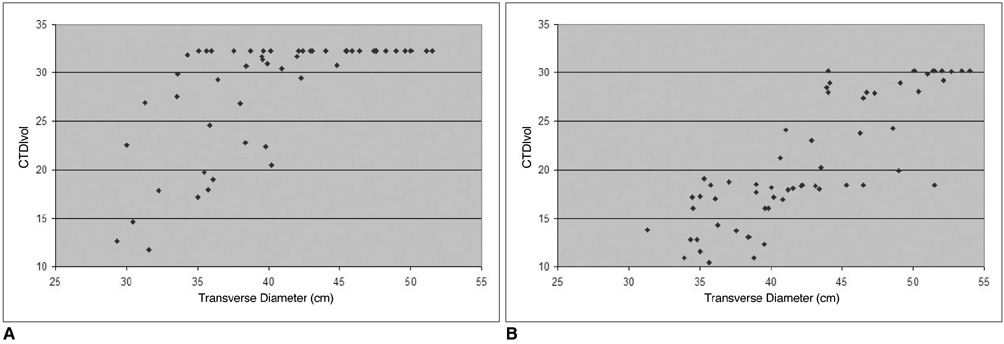
Fig. 1 Scatter plot of radiation dose (CTDIvol-y-axis) versus patient transverse diameter (cm) (x-axis) showing moderate correlation for chest CT performed by non-weight-adjusted automatic exposure control technique (correlation coefficient = 0.61) (A). Strong correlation (correlation coefficient = 0.83) was observed for chest CT performed by weight-adjusted automatic exposure control technique (B).
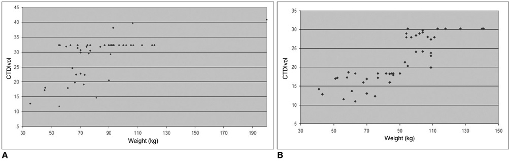
Fig. 2 Scatter plot graph of radiation dose (CTDIvol - y-axis) versus patient weight (kg) (x-axis) shows moderate correlation for chest CT performed by non-weight-adjusted automatic exposure control technique (correlation coefficient = 0.66) (A). Strong correlation (correlation coefficient = 0.83) was observed for chest CT performed by weight-adjusted automatic exposure control technique (B).
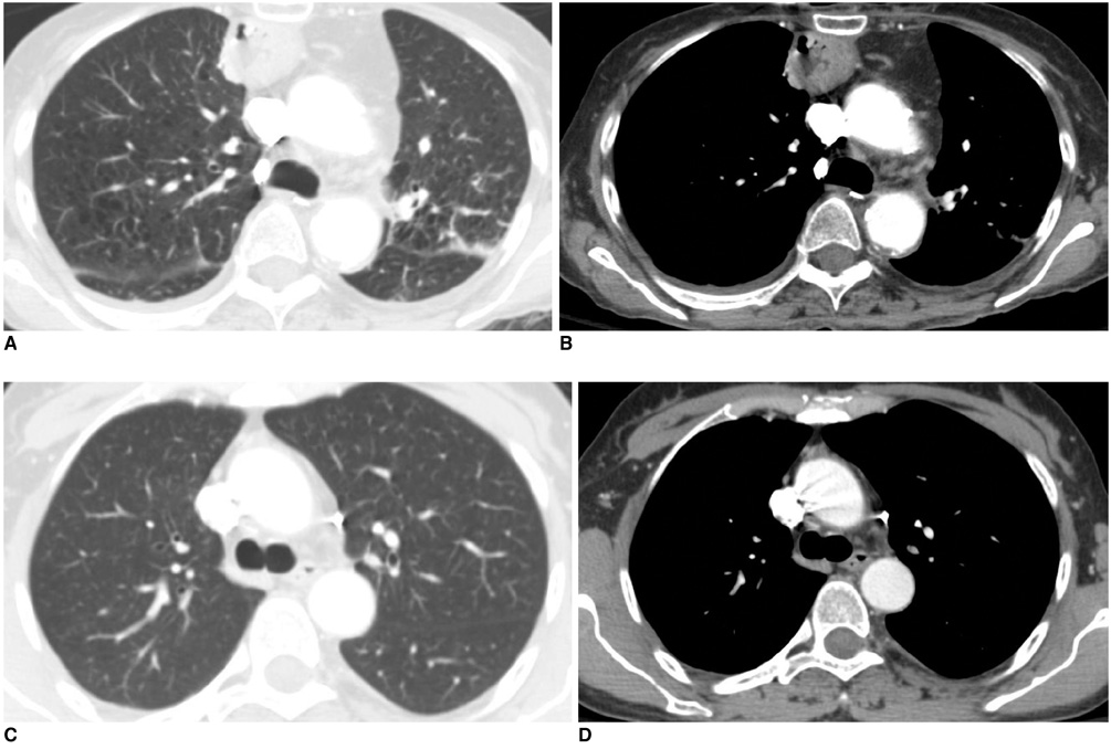
Fig. 3 2.5 mm thick transverse chest CT performed by weight-adapted automatic exposure control technique in lung (A) and mediastinal (B) windows in 76-year-old female (weight 43 kg, objective noise 8.1, and CTDIvol 12.8 mGy). Chest CT examination with non-weight-adjusted automatic exposure control technique in 43-year-old female (weight 45 kg, objective noise 8.9, CTDIvol 18.0 mGy) in lung (C) and mediastinal (D) windows. At lower radiation dose, weight-adjusted automatic exposure control protocol provides acceptable image noise and diagnostic acceptability.
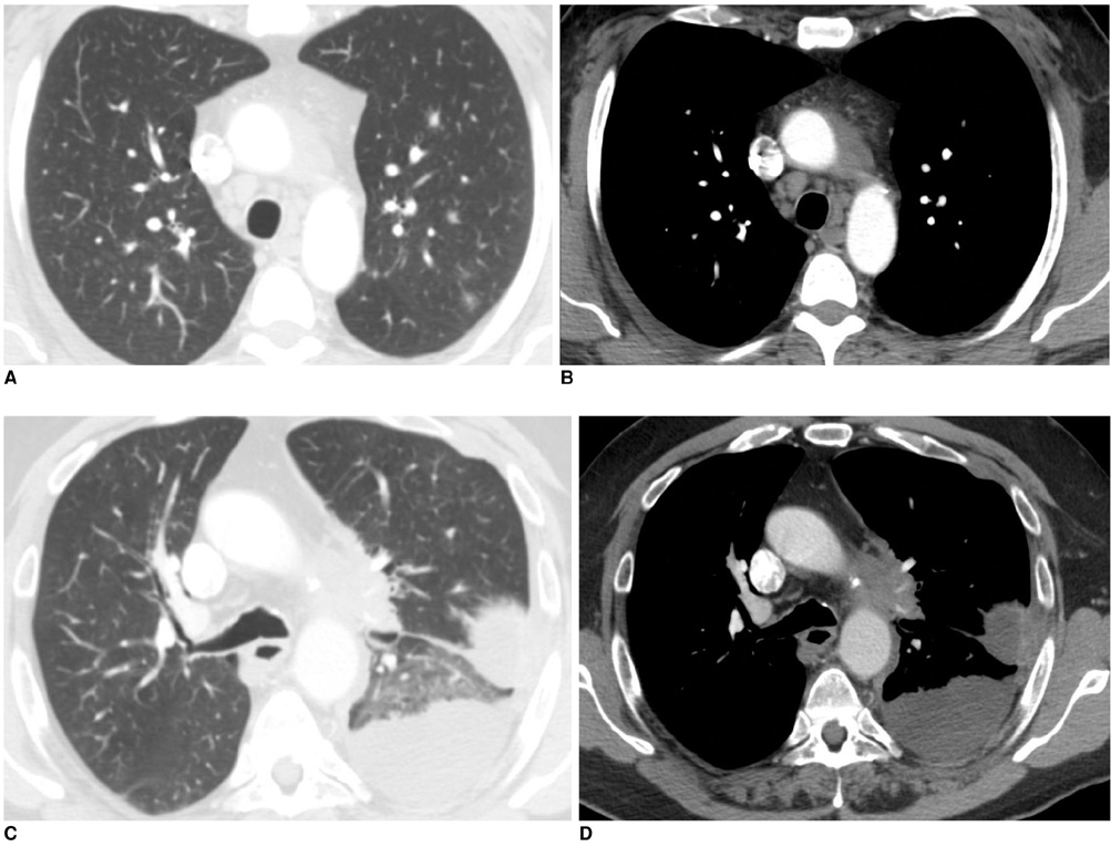
Fig. 4 2.5 mm thick transverse chest CT performed with weight-adapted automatic exposure control technique for lung (A) and mediastinal (B) windows in 46-year-old male (patient weight 107 kg, objective noise 10.5, and CTDIvol 28.0 mGy). Chest CT examinations using non-weight-adjusted automatic exposure control technique in 58-year-old male (patient weight 107 kg, objective noise 10.6, and CTDIvol 39.6 mGy) for lung (C) and mediastinal (D) windows. At lower radiation dose, weight-based automatic exposure control protocols provides acceptable image noise and diagnostic acceptability.
Cited by 1 articles
-
Estimation of Radiation Exposure of 128-Slice 4D-Perfusion CT for the Assessment of Tumor Vascularity
Dominik Ketelsen, Marius Horger, Markus Buchgeister, Michael Fenchel, Christoph Thomas, Nadine Boehringer, Maximilian Schulze, Ilias Tsiflikas, Claus D. Claussen, Martin Heuschmid
Korean J Radiol. 2010;11(5):547-552. doi: 10.3348/kjr.2010.11.5.547.
Reference
-
1. Mayo JR, Aldrich J, Muller NL. Fleischner Society. Radiation exposure at chest CT: a statement of the Fleischner Society. Radiology. 2003. 228:15–21.2. Mettler FA Jr, Wiest PW, Locken JA, Kelsey CA. CT scanning: patterns of use and dose. J Radiol Prot. 2000. 20:353–359.3. McCollough CH, Bruesewitz MR, Kofler JM Jr. CT dose reduction and dose management tools: overview of available options. Radiographics. 2006. 26:503–512.4. Kalra MK, Maher MM, Toth TL, Hamberg LM, Blake MA, Shepard JA, et al. Strategies for CT radiation dose optimization. Radiology. 2004. 230:619–628.5. Kalra MK, Maher MM, Kamath RS, Horiuchi T, Toth TL, Halpern EF, et al. Sixteen-detector row CT of abdomen and pelvis: study for optimization of Z-axis modulation technique performed in 153 patients. Radiology. 2004. 233:241–249.6. European guidelines on quality criteria for computed tomography. EUR 16262. Accessed on January 13, 2009. Available at: www.drs.dk/guidelines/ct/quality/download/eur16262.w51.7. Kalra MK, Rizzo S, Maher MM, Halpern EF, Toth TL, Shepard JA, et al. Chest CT performed with z-axis modulation: scanning protocol and radiation dose. Radiology. 2005. 237:303–308.8. Kubo T, Lin PJ, Stiller W, Takahashi M, Kauczor HU, Ohno Y, et al. Radiation dose reduction in chest CT: a review. AJR Am J Roentgenol. 2008. 190:335–343.9. Prasad SR, Wittram C, Shepard JA, McLoud T, Rhea J. Standard-dose and 50%-reduced-dose chest CT: comparing the effect on image quality. AJR Am J Roentgenol. 2002. 179:461–465.10. Karabulut N, Ariyürek M. Low dose CT: practices and strategies of radiologists in university hospitals. Diagn Interv Radiol. 2006. 12:3–8.11. Mulkens TH, Bellinck P, Baeyaert M, Ghysen D, Van Dijck X, Mussen E, et al. Use of an automatic exposure control mechanism for dose optimization in multi-detector row CT examinations: clinical evaluation. Radiology. 2005. 237:213–223.12. Zhu X, Yu J, Huang Z. Low-dose chest CT: optimizing radiation protection for patients. AJR Am J Roentgenol. 2004. 183:809–816.13. Mayo JR, Kim KI, MacDonald SL, Johkoh T, Kavanagh P, Coxson HO, et al. Reduced radiation dose helical chest CT: effect on reader evaluation of structures and lung findings. Radiology. 2004. 232:749–756.
- Full Text Links
-
- Actions
-
Cited
- CITED
-
- Close
- Share
-
- Similar articles
-
- In-Plane Shielding for CT: Effect of Off-Centering, Automatic Exposure Control and Shield-to-Surface Distance
- The Dose and Risk Reduction from Adoption of Automatic mA Control in 4D CT Scans
- Radiation exposure from Chest CT: Issues and Strategies
- CT radiation dose and radiation reduction strategies
- Attenuation-Based Automatic Kilovoltage Selection and Sinogram-Affirmed Iterative Reconstruction: Effects on Radiation Exposure and Image Quality of Portal-Phase Liver CT
