In-Plane Shielding for CT: Effect of Off-Centering, Automatic Exposure Control and Shield-to-Surface Distance
- Affiliations
-
- 1Department of Radiology, Massachusetts General Hospital, Boston MA 02114, USA. mkalra@partners.org
- KMID: 1088727
- DOI: http://doi.org/10.3348/kjr.2009.10.2.156
Abstract
OBJECTIVE
To assess effects of off-centering, automatic exposure control, and padding on attenuation values, noise, and radiation dose when using in-plane bismuth-based shields for CT scanning.
MATERIALS AND METHODS
A 30 cm anthropomorphic chest phantom was scanned on a 64-multidetector CT, with the center of the phantom aligned to the gantry isocenter. Scanning was repeated after placing a bismuth breast shield on the anterior surface with no gap and with 1, 2, and 6 cm of padding between the shield and the phantom surface. The "shielded" phantom was also scanned with combined modulation and off-centering of the phantom at 2 cm, 4 cm and 6 cm below the gantry isocenter. CT numbers, noise, and surface radiation dose were measured. The data were analyzed using an analysis of variance.
RESULTS
The in-plane shield was not associated with any significant increment for the surface dose or CT dose index volume, which was achieved by comparing the radiation dose measured by combined modulation technique to the fixed mAs (p > 0.05). Irrespective of the gap or the surface CT numbers, surface noise increased to a larger extent compared to Hounsfield unit (HU) (0-6 cm, 26-55%) and noise (0-6 cm, 30-40%) in the center. With off-centering, in-plane shielding devices are associated with less dose savings, although dose reduction was still higher than in the absence of shielding (0 cm off-center, 90% dose reduction; 2 cm, 61%) (p < 0.0001). Streak artifacts were noted at 0 cm and 1 cm gaps but not at 2 cm and 6 cm gaps of shielding to the surface distances.
CONCLUSION
In-plane shields are associated with greater image noise, artifactually increased attenuation values, and streak artifacts. However, shields reduce radiation dose regardless of the extent of off-centering. Automatic exposure control did not increase radiation dose when using a shield.
MeSH Terms
Figure
-
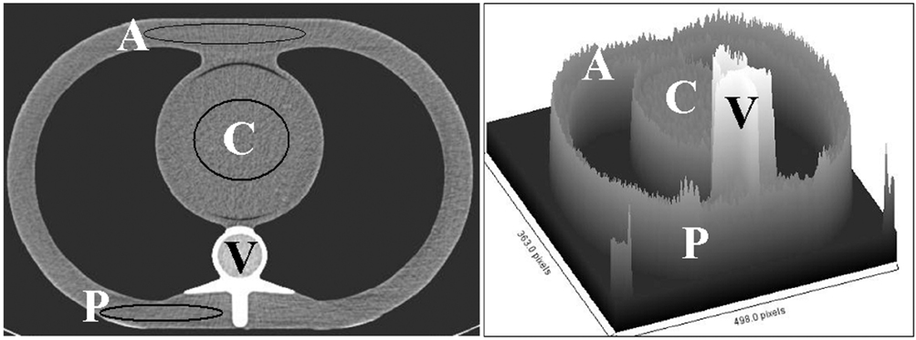
Fig. 1 Transverse CT image and surface plot of phantom without shield. Three regions of interest were drawn on CT images at same level and position in anterior (A), central (C) and posterior (P) regions of phantom. As expected, vertebra has highest CT numbers on surface plot image.

Fig. 2 Original transverse CT images of phantom (*) acquired at 0 cm (Ao), 1 cm (Bo), 2 cm (Co), and 6 cm (Do) offsets (black arrows) and their corresponding CT numbers' contour maps (A, B, C, D). Compared to image acquired with 6 cm offset (D), images with 0-2 cm offsets have considerably higher CT numbers and lighter shade of gray on surface plots (A, B, C). Also note difference in CT numbers in anterior and posterior aspects of phantom.
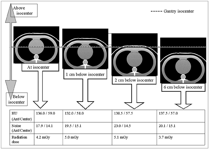
Fig. 3 Off-centering of shielded phantom below gantry isocenter did not cause any substantial change in CT numbers (HU ant/center in anterior or central regions of the phantom), noise, and average surface radiation dose compared to scanning with appropriate phantom centering.
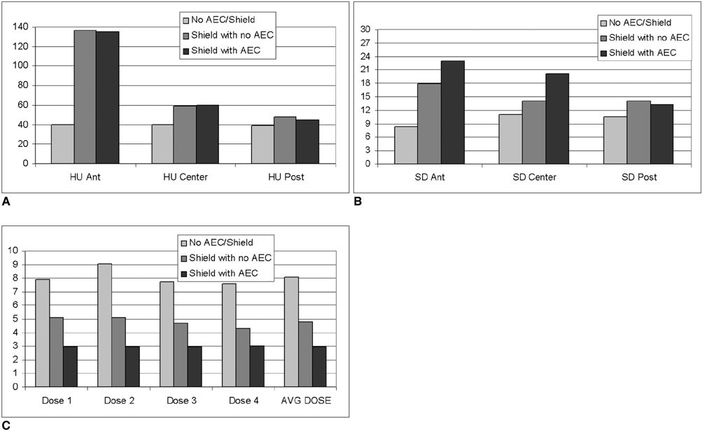
Fig. 4 Combined modulation technique (AEC) and plane shielding. A. Compared to images acquired without shield, CT numbers in anterior (HU ant) and central (HU center) regions of phantom are substantially higher with shield, with and without combined modulation, whereas CT numbers are similar in posterior region (HU post) of phantom. B. Compared to images acquired without shield, image noise in anterior (SD ant) and central (SD center) regions of phantom are substantially higher with shield, with and without combined modulation, whereas image noise is similar in posterior region (SD post) of phantom. In particular, combined modulation scanning was associated with greater image noise compared to fixed tube current scanning when phantom was scanned with shield for both techniques. C. Comparison of images acquired with or without shield, with both conditions having constant fixed tube current, combined modulation technique resulted in significantly lower individual (doses 1, 2, 3, 4) and average surface radiation doses (AVG DOSE).
Cited by 1 articles
-
Estimation of Radiation Exposure of 128-Slice 4D-Perfusion CT for the Assessment of Tumor Vascularity
Dominik Ketelsen, Marius Horger, Markus Buchgeister, Michael Fenchel, Christoph Thomas, Nadine Boehringer, Maximilian Schulze, Ilias Tsiflikas, Claus D. Claussen, Martin Heuschmid
Korean J Radiol. 2010;11(5):547-552. doi: 10.3348/kjr.2010.11.5.547.
Reference
-
1. Kalra MK, Maher MM, Toth TL, Hamberg LM, Blake MA, Shepard JA, et al. Strategies for CT radiation dose optimization. Radiology. 2004. 230:619–628.2. Li J, Udayasankar UK, Toth TL, Seamans J, Small WC, Kalra MK. Automatic patient centering for MDCT: effect on radiation dose. AJR Am J Roentgenol. 2007. 188:547–552.3. Mukundan S Jr, Wang PI, Frush DP, Yoshizumi T, Marcus J, Kloeblen E, et al. MOSFET dosimetry for radiation dose assessment of bismuth shielding of the eye in children. AJR Am J Roentgenol. 2007. 188:1648–1650.4. Colombo P, Pedroli G, Nicoloso M, Re S, Valvassori L, Vanzulli A. Evaluation of the efficacy of a bismuth shield during CT examinations. Radiol Med (Torino). 2004. 108:560–568.5. Hopper KD, Neuman JD, King SH, Kunselman AR. Radioprotection to the eye during CT scanning. AJNR Am J Neuroradiol. 2001. 22:1194–1198.6. Hopper KD. Orbital, thyroid, and breast superficial radiation shielding for patients undergoing diagnostic CT. Semin Ultrasound CT MR. 2002. 23:423–427.7. Dauer LT, Casciotta KA, Erdi YE, Rothenberg LN. Radiation dose reduction at a price: the effectiveness of a male gonadal shield during helical CT scans. BMC Med Imaging. 2007. 7:5.8. Primak AN, McCollough CH, Bruesewitz MR, Zhang J, Fletcher JG. Relationship between noise, dose, and pitch in cardiac multi-detector row CT. Radiographics. 2006. 26:1785–1794.9. Yoshizumi TT, Goodman PC, Frush DP, Nguyen G, Toncheva G, Sarder M, et al. Validation of metal oxide semiconductor field effect transistor technology for organ dose assessment during CT: comparison with thermoluminescent dosimetry. AJR Am J Roentgenol. 2007. 188:1332–1336.10. Rizzo S, Kalra M, Schmidt B, Dalal T, Suess C, Flohr T, et al. Comparison of angular and combined automatic tube current modulation techniques with constant tube current CT of the abdomen and pelvis. AJR Am J Roentgenol. 2006. 186:673–679.11. Fricke BL, Donnelly LF, Frush DP, Yoshizumi T, Varchena V, Poe SA, et al. In-plane bismuth breast shields for pediatric CT: effects on radiation dose and image quality using experimental and clinical data. AJR Am J Roentgenol. 2003. 180:407–411.12. McLaughlin DJ, Mooney RB. Dose reduction to radiosensitive tissues in CT. Do commercially available shields meet the users' needs? Clin Radiol. 2004. 59:446–450.13. Geleijns J, Salvadó Artells M, Veldkamp WJ, López Tortosa M, Calzado Cantera A. Quantitative assessment of selective in-plane shielding of tissues in computed tomography through evaluation of absorbed dose and image quality. Eur Radiol. 2006. 16:2334–2340.
- Full Text Links
-
- Actions
-
Cited
- CITED
-
- Close
- Share
-
- Similar articles
-
- A Study on Dose Distribution around Fletcher-Suit Colpostat Containing 137Cs Source
- A New Shielding Curtain for Protection of Intraoperative Radiation During Minimally Invasive Spine Surgery
- Evaluations of the Space Dose and Dose Reductions in Patients and Practitioners by Using the C-arm X-ray Tube Shielding Devices Developed in Our Laboratory
- Reviews of Radiation Protection and Shielding for Computed Tomography in Foreign Countries
- A Study on Dose Distribution outside Co-0 gamma Ray and 10MV X Ray Fields
