J Korean Med Sci.
2004 Apr;19(2):159-166. 10.3346/jkms.2004.19.2.159.
Radiation exposure from Chest CT: Issues and Strategies
- Affiliations
-
- 1Department of Radiology, Massachusetts General Hospital and Harvard Medical School, USA. mkalra@partners.org
- 2Department of Pulmonary and Critical Care Medicine, Massachusetts General Hospital and Harvard Medical School, USA.
- KMID: 1733471
- DOI: http://doi.org/10.3346/jkms.2004.19.2.159
Abstract
- Concerns have been raised over alleged overuse of CT scanning and inappropriate selection of scanning methods, all of which expose patients to unnecessary radiation. Thus, it is important to identify clinical situations in which techniques with lower radiation dose such as plain radiography or no radiation such as MRI and occasionally ultrasonography can be chosen over CT scanning. This article proposes the arguments for radiation dose reduction in CT scanning of the chest and discusses recommended practices and studies that address means of reducing radiation exposure associated with CT scanning of the chest.
Keyword
MeSH Terms
Figure
-
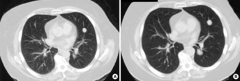
Fig. 1 Low radiation dose images can also give diagnostic quality images. Transverse CT images reveal multiple metastatic nodules in a 64-yr-old man with colon cancer who underwent a standard radiation dose CT (224 mAs) (A) and follow-up CT with 50% reduction in radiation dose (112 mAs) (B).
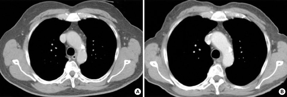
Fig. 2 Technology can aid in radiation dose reduction. Transverse CT image (224 mAs) (A) of a 44-yr-old man with chronic cough acquired with conventional scanning technique is similar to CT image (112 mAs) (B) acquired with automatic tube current modulation technique (at 50% reduction in radiation dose) in terms of diagnostic quality.
Reference
-
1. EUR 16262 Commission of the European Community. Report EUR 16262 EN. European guidelines on quality criteria for computed tomography. 1999.2. UNSCEAR 2000. The United Nations Scientific Committee on the Effects of Atomic Radiation. Health Phys. 2000. 79:314.3. Tack Group on Control of Radiation Dose in Computed Tomography. Managing patient dose in Computed Tomography. A report of the International Commission on Radiological Protection. Ann ICRP. 2000. 30:7–45.4. Gray JE. Janower ML, Linton OW, editors. Safety (risk) of diagnostic radiology exposures. Radiation risk: a primer. 1996. Reston, VA: American College of Radiology;15–17.5. Wagner LK, Eifel PJ, Geise RA. Potential biological effects following high X-ray dose interventional procedures. J Vasc Interv Radiol. 1994. 5:71–84.
Article6. Huda W, Peters KR. Radiation-induced temporary epilation after a neuroradiologically guided embolization procedure. Radiology. 1994. 193:642–644.
Article7. Koenig TR, Wolff D, Mettler FA, Wagner LK. Skin injuries from fluoroscopically guided procedures: part 1, characteristics of radiation injury. AJR Am J Roentgenol. 2001. 177:3–11.8. Koenig TR, Mettler FA, Wagner LK. Skin injuries from fluoroscopically guided procedures: part 2, review of 73 cases and recommendations for minimizing dose delivered to patient. AJR Am J Roentgenol. 2001. 177:13–20.9. IARC Monographs on the evaluation of carcinogenic risk to humans. Ionizing radiation, part 1: X- and gamma (γ)-radiation, and neutrons. 2000. Vol. 75. Lyons, France: International Agency for Research on Cancer (IARC).10. Diederich S, Lenzen H. Radiation exposure associated with imaging of the chest: comparison of different radiographic and computed tomography techniques. Cancer. 2000. 89:Suppl 11. 2457–2460.11. ICRP. Recommendations of the International Commission on Radiological Protection (Publication 60). 1991. Oxford: Pergamon Press.12. Wiest PW, Locken JA, Heintz PH, Mettler FA Jr. CT scanning: a major source of radiation exposure. Semin Ultrasound CT MR. 2002. 23:402–410.
Article13. Kalra MK, Prasad S, Saini S, Blake MA, Varghese J, Halpern EF, Thrall JH, Rhea JT. Clinical Comparison of Standard-Dose and 50% Reduced-Dose Abdominal CT: Effect on Image Quality. AJR Am J Roentgenol. 2002. 179:1101–1106.14. Slovis TL. CT and computed radiography: The pictures are great, but is the radiation dose greater than required? AJR Am J Roentgenol. 2002. 179:39–41.
Article15. Karabulut N, Martin DR, Yang M, Tallaksen RJ. MR Imaging of the Chest using a Contrast-enhanced breath-hold modified three-dimensional Gradient-Echo technique: comparison with two-dimensional Gradient-Echo technique and multidetector CT. AJR Am J Roentgenol. 2002. 179:1225–1233.
Article16. Johkoh T, Muller NL, Nakamura H. Multidetector spiral high-resolution computed tomography of the lungs: distribution of findings on coronal image reconstructions. J Thorac Imaging. 2002. 17:291–305.
Article17. Prasad SR, Wittram C, Shepard JA, McLoud T, Rhea J. Standard-dose and 50%-reduced-dose chest CT: comparing the effect on image quality. AJR Am J Roentgenol. 2002. 179:461–465.18. Prokop M. Optimizing dosage in thoracic computerized tomography. Radiologe. 2001. 41:269–278.19. Donnelly LF, Emery KH, Brody AS, Laor T, Gylys-Morin VM, Anton CG, Thomas SR, Frush DP. Minimizing Radiation Dose for Pediatric Body Applications of Single-Detector Helical CT: Strategies at a Large Children's Hospital. AJR Am J Roentgenol. 2001. 176:303–306.20. Lucaya J, Piqueras J, Garcia-Pena P, Enriquez G, Garcia-Macias M, Sotil J. Low-dose high-resolution CT of the chest in children and young adults: dose, cooperation, artifact incidence, and image quality. AJR Am J Roentgenol. 2000. 175:985–992.21. Wildberger JE, Mahnken AH, Schmitz-Rode T, Flohr T, Stargardt A, Haage P, Schaller S, Gunther RW. Individually adapted examination protocols for reduction of radiation exposure in chest CT. Invest Radiol. 2001. 36:604–611.
Article22. Hidajat N, Schroder RJ, Vogl T, Schedel H, Felix R. The efficacy of lead shielding in patient dosage reduction in computed tomography. Rofo Fortschr Geb Rontgenstr Neuen Bildgeb Verfahr. 1996. 165:462–465.23. Hopper KD, King SH, Lobell ME, TenHave TR, Weaver JS. The breast: in-plane x-ray protection during diagnostic thoracic CT--shielding with bismuth radioprotective garments. Radiology. 1997. 205:853–858.
Article24. Hopper KD. Orbital, thyroid, and breast superficial radiation shielding for patients undergoing diagnostic CT. Semin Ultrasound CT MR. 2002. 23:423–427.
Article25. Huda W, Scalzetti EM, Roskopf M. Effective doses to patients undergoing thoracic computed tomography examinations. Med Phys. 2000. 27:838–844.
Article26. Takahashi M, Maguire WM, Ashtari M, Khan A, Papp Z, Alberico R, Campbell W, Eacobacci T, Herman PG. Low-dose spiral computed tomography of the thorax: comparison with the standard-dose technique. Invest Radiol. 1998. 33:68–73.27. Diederich S, Lenzen H, Puskas Z, Koch AT, Yelbuz TM, Eameri M, Roos N, Peters PE. Low dose computerized tomography of the thorax. Experimental and clinical studies. Radiologe. 1996. 36:475–482.28. Huda W, Ravenel JG, Scalzetti EM. How do radiographic techniques affect image quality and patient doses in CT? Semin Ultrasound CT MR. 2002. 23:411–422.
Article29. Ambrosino MM, Genieser NB, Roche KJ, Kaul A, Lawrence RM. Feasibility of high-resolution, low-dose chest CT in evaluating the pediatric chest. Pediatr Radiol. 1994. 24:6–10.
Article30. Zwirewich CV, Mayo JR, Muller NL. Low-dose high-resolution CT of lung parenchyma. Radiology. 1991. 180:413–417.
Article31. Mayo JR, Jackson SA, Muller NL. High-resolution CT of the chest: radiation dose. AJR Am J Roentgenol. 1993. 160:479–481.
Article32. Lee KS, Primack SL, Staples CA, Mayo JR, Aldrich JE, Muller NL. Chronic infiltrative lung disease: comparison of diagnostic accuracies of radiography and low- and conventional-dose thin-section CT. Radiology. 1994. 191:669–673.
Article33. Reuter M, Oppermann HC, Ankermann T, Biederer J, Heller M. High-resolution computed tomography of the lungs in pediatric patients. Rofo Fortschr Geb Rontgenstr Neuen Bildgeb Verfahr. 2002. 174:684–695.
Article34. Diederich S, Wormanns D, Heindel W. Low-dose CT: new tool for screening lung cancer? Eur Radiol. 2001. 11:1916–1924.
Article35. Oguchi K, Sone S, Kiyono K, Takashima S, Maruyama Y, Hasegawa M, Feng L. Optimal tube current for lung cancer screening with low-dose spiral CT. Acta Radiol. 2000. 41:352–356.
Article36. Kaneko M, Kusumoto M, Kobayashi T, Moriyama N, Naruke T, Ohmatsu H, Kakinuma R, Eguchi K, Nishiyama H, Matsui E. Computed tomography screening for lung carcinoma in Japan. Cancer. 2000. 89:2485–2488.
Article37. Michel JL, Reynier C, Avy G, Bard JJ, Gabrillargues D, Catilina P. An assessment of low-dose high resolution CT in the detection of benign asbestos-related pleural abnormalities. J Radiol. 2001. 82:922–923.38. Lederle FA, Nichol KL, Parenti CM. Bronchoscopy to evaluate hmoptysis in older men with nonsuspicious chest roentgenograms. Chest. 1989. 95:1043–1047.39. Haponik EF, Britt EJ, Smith PL, Bleecker ER. Computed chest tomography in the evaluation of hemoptysis. Impact on diagnosis and treatment. Chest. 1987. 91:80–85.40. Set PA, Flower CD, Smith IE, Chan AP, Twentyman OP, Shneerson JM. Hemoptysis: comparative study of the role of CT and fiberoptic bronchoscopy. Radiology. 1993. 189:677–680.
Article41. Lim DJ, Carter MF. Computerized tomography in the preoperative staging for pulmonary metastases in patients with renal cell carcinoma. J Urol. 1993. 150:1112–1114.
Article42. See WA, Hoxie L. Chest staging in testis cancer patients: imaging modality selection based upon risk assessment as determined by abdominal computerized tomography scan results. J Urol. 1993. 150:874–878.
Article43. Gartenschlager M, Schweden F, Gast K, Westermeier T, Kauczor H, von Zitzewitz H, Thelen M. Pulmonary nodules: detection with low-dose vs conventional-dose spiral CT. Eur Radiol. 1998. 8:609–614.44. Diederich S, Lenzen H, Windmann R, Puskas Z, Yelbuz TM, Henneken S, Klaiber T, Eameri M, Roos N, Peters PE. Pulmonary nodules: experimental and clinical studies at low-dose CT. Radiology. 1999. 213:289–298.
Article45. Ravenel JG, Scalzetti EM, Huda W, Garrisi W. Radiation exposure and image quality in chest CT examinations. AJR Am J Roentgenol. 2001. 177:279–284.
Article46. Toth TL, Bromberg NB, Pan TS, Rabe J, Woloschek SJ, Li J, Seidenschnur GE. A dose reduction x-ray beam positioning system for high-speed multislice CT scanners. Med Phys. 2000. 27:2659–2668.
Article47. Fox SH, Toth T. Dose reduction on GE CT scanners. Pediatr Radiol. 2002. 32:718–723.
Article48. Kachelriess M, Watzke O, Kalender WA. Generalized multi-dimensional adaptive filtering for conventional and spiral single-slice, multi-slice, and cone-beam CT. Med Phys. 2001. 28:475–490.49. Greess H, Wolf H, Baum U, Lell M, Pirkl M, Kalender W, Bautz WA. Dose reduction in computed tomography by attenuation-based on-line modulation of tube current: evaluation of six anatomical regions. Eur Radiol. 2000. 10:391–394.
Article50. Itoh S, Koyama S, Ikeda M, Ozaki M, Sawaki A, Iwano S, Ishigaki T. Further reduction of radiation dose in helical CT for lung cancer screening using small tube current and a newly designed filter. J Thorac Imaging. 2001. 16:81–88.
Article51. Kalra MK, Wittram C, Maher MM, Sharma A, Avinash GB, Karau K, Toth TL, Halpern E, Saini S, Shepard JA. Can Noise Reduction Filters Improve Low Radiation Dose Chest CT Images? - pilot study. Radiology. 2003. 228:257–264.52. Kalra MK, Maher MM, Sahani DV, Blake MA, Hahn PF, Avinash GB, Toth TL, Halpern E, Saini S. Low-dose CT of the abdomen: Evaluation of image improvement with use of noise reduction filters- pilot study. Radiology. 2003. 228:251–256.53. Kalra MK, Maher MM, Lucey BC, Blake M, Karau K, Saini S. Lesion detection on reduced radiation dose CT images processed with noise reduction filters (abstract). Radiology. 2002. 645.54. Frush DP, Slack CC, Hollingsworth CL, Bisset GS, Donnelly LF, Hsieh J, Lavin-Wensell T, Mayo JR. Computer-simulated radiation dose reduction for abdominal multidetector CT of pediatric patients. AJR Am J Roentgenol. 2002. 179:1107–1113.
Article55. Mayo JR, Whittall KP, Leung AN, Hartman TE, Park CS, Primack SL, Chambers GK, Limkeman MK, Toth TL, Fox SH. Simulated dose reduction in conventional chest CT: validation study. Radiology. 1997. 202:453–457.
Article56. Kersjes W, Mayer E, Buchenroth M, Schunk K, Fouda N, Cagil H. Diagnosis of pulmonary metastases with turbo-SE MR imaging. Eur Radiol. 1997. 7:1190–1194.
Article57. Thompson BH, Stanford W. MR imaging of pulmonary and mediastinal malignancies. Magn Reson Imaging Clin N Am. 2000. 8:729–739.58. Muller NL. Computed tomography and magnetic resonance imaging: past, present and future. Eur Respir J Suppl. 2002. 35:3s–12s.
Article59. Fattori R, Nienaber CA. MRI of acute and chronic aortic pathology: pre-operative and postoperative evaluation. J Magn Reson Imaging. 1999. 10:741–750.
Article60. Kangarloo H. Chest MRI in children. Radiol Clin North Am. 1988. 26:263–275.61. Kauczor HU, Heussel CP, Schreiber WG, Kreitner KF. New developments in MRI of the thorax. Radiologe. 2001. 41:279–287.62. Luo L, Hierholzer J, Bittner RC, Chen J, Huang L. Magnetic resonance imaging in distinguishing malignant from benign pleural disease. Chin Med J (Engl). 2001. 114:645–649.63. Heelan RT, Rusch VW, Begg CB, Panicek DM, Caravelli JF, Eisen C. Staging of malignant pleural mesothelioma: comparison of CT and MR imaging. AJR Am J Roentgenol. 1999. 172:1039–1047.
Article64. Gavelli G, Canini R, Bertaccini P, Battista G, Bna C, Fattori R. Traumatic injuries: imaging of thoracic injuries. Eur Radiol. 2002. 12:1273–1294.
Article65. Yang PC. Ultrasound-guided transthoracic biopsy of the chest. Radiol Clin North Am. 2000. 38:323–343.
Article66. Mathis G, Gehmacher O. Lung and pleural ultrasound. Schweiz RundschMed Prax. 2001. 90:681–686.67. Madan A, van Rooij WJ, Verpalen MC. Sonographically guided needle biopsy in peripheral thoracic masses: results in 50 patients. Rofo Fortschr Geb Rontgenstr Neuen Bildgeb Verfahr. 1994. 160:75–77.
Article68. Sheth S, Hamper UM, Stanley DB, Wheeler JH, Smith PA. US guidance for thoracic biopsy: a valuable alternative to CT. Radiology. 1999. 210:721–726.
Article69. Catalano MF, Rosenblatt ML, Chak A, Sivak MV Jr, Scheiman J, Gress F. Endoscopic ultrasound-guided fine needle aspiration in the diagnosis of mediastinal masses of unknown origin. Am J Gastroenterol. 2002. 97:2559–2565.
Article
- Full Text Links
-
- Actions
-
Cited
- CITED
-
- Close
- Share
-
- Similar articles
-
- CT radiation dose and radiation reduction strategies
- General Principles of Radiation Protection in Fields of Diagnostic Medical Exposure
- Pediatric CT: Understanding of Radiation Dose and Optimization of Imaging Techniques
- The health effects of low-dose radiation exposure
- A study on radiation exposure dose at brain CT
