Sixteen Cases of Sclerosing Hemangioma of the Lung Including Unusual Presentations
- Affiliations
-
- 1Department of Pathology, Sungkyunkwan University School of Medicine, Samsung Medical Center, Seoul, Korea. jhhan@smc.samsung.co.kr
- 2Department of Thoracic Surgery, Division of Pulmonology and Critical Care Medicine, Sungkyunkwan University School of Medicine, Samsung Medical Center, Seoul, Korea.
- 3Department of Internal Medicine, Sungkyunkwan University School of Medicine, Samsung Medical Center, Seoul, Korea.
- KMID: 1786811
- DOI: http://doi.org/10.3346/jkms.2004.19.3.352
Abstract
- Sclerosing hemangiomas (SH) of the lung are uncommon tumors and are thought to be benign. However, the biologic behavior of this tumor has not yet been characterized adequately. The clinicopathologic features were reviewed and analyzed for 16 cases of SH. The age of the patients ranged from 37 to 73 yr (mean 50.6 yr). There were fifteen female and one male patient. The SH located at the intraparenchyme in 14 cases, the interlobar fissure in one case and the visceral pleura in one case. The size of SH ranged from 0.3 cm to 8 cm (mean 2.6 cm). There were five unusual presentations of SH including a case having two SH with multiple nodules of atypical adenomatous hyperplasia in the same lobe, a case showing adenocarcinomalike area within the SH, a case showing one peribronchial lymph node metastasis (N1 nodal stage) with location of interlobar major fissure, a case showing alveolar adenoma-like area within the SH, and one case with a large visceral pleural-based pedunculated mass presenting as mediastinal mass. All patients were alive and well without recurrence at the last follow up. Here, we reviewed previously published literatures and discussed the histogenesis of SH.
Keyword
MeSH Terms
Figure
-
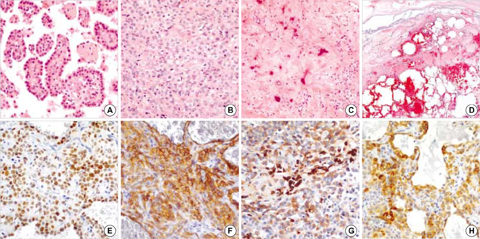
Fig. 1 Four major histologic patterns and immunohistochemistry of the sclerosing hemangiomas. Sclerosing hemangiomas show papillary (A), solid (B), sclerotic (C), and hemorrhagic (D) patterns. Sclerosing hemangioma consists of lining cuboidal cells (A) and stromal round cells (B) (H&E stain: A, B ×200; C, ×100; D, ×1). Immunohistochemical stain shows that both lining cells and round cells are positive for TTF-1 (E) and EMA (F), and CD56 (G). The pancytokeratin (H) reacts with the lining cells and focally reacts with round cells. (E to H, ×200).

Fig. 2 Unusual presentations of the sclerosing hemangiomas. Two sclerosing hemangiomas (A, B) with atypical alveolar hyperplasia-like nodule (C) in the background lung parenchyme (H&E stain: A, B, ×1; C, ×200). Atypical alveolar hyperplasia-like nodule shows that the lining cells and some stromal cells are positive for TTF-1 (D) immunostaining (×400). (E) One sclerosing hemangioma with lymph node metastasis (H&E stain, ×200). (F) One sclerosing hemangioma with an alveolar adenoma-like area in the upper half (H&E stain, ×100). (G) One sclerosing hemangioma with adenocarcinoma-like area (H&E stain, ×100).
Cited by 1 articles
-
Solitary Capillary Hemangioma of the Lung: A Report of Two Cases
Hyun-Woo Lee, Soomin Ahn, Young Mog Shim, Yong Soo Choi, Kyung-Soo Lee, Joungho Han
J Lung Cancer. 2012;11(2):102-104. doi: 10.6058/jlc.2012.11.2.102.
Reference
-
1. Liebow AA, Hubbell DS. Sclerosing hemangioma (histiocytoma, xanthoma) of the lung. Cancer. 1956. 9:53–75.
Article2. Huszar M, Suster S, Herczeg E, Geiger B. Sclerosing hemangioma of the lung. Immunohistochemical demonstration of mesenchymal origin using antibodies to tissue-specific intermediate filaments. Cancer. 1986. 58:2422–2427.
Article3. Katzenstein AL, Weise DL, Fulling K, Battifora H. So-called sclerosing hemangioma of the lung. Evidence for mesothelial origin. Am J Surg Pathol. 1983. 7:3–14.4. Xu HM, Li WH, Hou N, Zhang SG, Li HF, Wang SQ, Yu ZY, Li ZJ, Zeng MY, Zhu GM. Neuroendocrine differentiation in 32 cases of so-called sclerosing hemangioma of the lung: identified by immunohistochemical and ultrastructural study. Am J Surg Pathol. 1997. 21:1013–1022.
Article5. Chan AC, Chan JK. Pulmonary sclerosing hemangioma consistently expresses thyroid transcription factor-1 (TTF-1): a new clue to its histogenesis. Am J Surg Pathol. 2000. 24:1531–1536.6. Devouassoux-Shisheboran M, Hayashi T, Linnoila RI, Koss MN, Travis WD. A clinicopathologic study of 100 cases of pulmonary sclerosing hemangioma with immunohistochemical studies: TTF-1 is expressed in both round and surface cells, suggesting an origin from primitive respiratory epithelium. Am J Surg Pathol. 2000. 24:906–916.7. World Health Organization. Histological Typing of Lung Tumors. 1981. Geneva: World Health Organization.8. Travis WD, Colby TV, Corrin B, Brambilla E. Histological Typing of Lung and Pleural Tumors. 1999. Berlin: Springer.9. Rodriguez-Soto J, Colby TV, Rouse RV. A critical examination of the immunophenotype of pulmonary sclerosing hemangioma. Am J Surg Pathol. 2000. 24:442–450.10. Stahlman MT, Gray ME, Whitsett JA. Expression of thyroid transcription factor-1 (TTF-1) in fetal and neonatal human lung. J Histochem Cytochem. 1996. 44:673–678.11. Khoor A, Whitsett JA, Stahlman MT, Olson SJ, Cagle PT. Utility of surfactant protein B precursor and thyroid transcription factor 1 in differentiating adenocarcinoma of the lung from malignant mesothelioma. Hum Pathol. 1999. 30:695–700.
Article12. Lin D, Zou S, Lu N, Liu X, Wen P, Li L. Thyroid transcription factor-1 in the histogenesis of plumonary sclerosing hemangioma. Zhonghua Zhong Liu Za Zhi. 2002. 24:384–387.13. Tanaka I, Inoue M, Matsui Y, Oritsu S, Akiyama O, Takemura T, Fujiwara M, Kodama T, Shimosato Y. A case of pneumocytoma (so-called sclerosing hemangioma) with lymph node metastasis. Jpn J Clin Oncol. 1986. 16:77–86.14. Spencer H, Nambu S. Sclerosing haemangiomas of the lung. Histopathology. 1986. 10:477–487.
Article15. Chung KY, Kim KD, Lim SH, Shin DH. A case of pneumocytoma (sclerosing hemangioma) with lymph node metastasis: a case report. Korean J Thorac Cardiovasc Surg. 1997. 30:548–551.16. Chen CS. Inflammatory pseudotumor and lung adenoma. Chin Med J (Engl). 1978. 4:297–298.17. Nicholson AG, Magkou C, Snead D, Vohra HA, Sheppard MN, Goldstraw P, Beddow E, Hansell DM, Travis WD, Corrin B. Unusual sclerosing haemangiomas and sclerosing haemangioma-like lesions, and the value of TTF-1 in making the diagnosis. Histopathology. 2002. 41:404–413.
Article18. Yano M, Yamakawa Y, Kiriyama M, Hara M, Murase T. Sclerosing hemangioma with metastases to multiple nodal stations. Ann Thorac Surg. 2002. 73:981–983.
Article19. Miyagawa-Hayashino A, Tazelaar HD, Langel DJ, Colby TV. Pulmonary sclerosing hemangioma with lymph node metastases: report of 4 cases. Arch Pathol Lab Med. 2003. 127:321–325.20. Kim KH, Sul HJ, Kang DY. Sclerosing hemangioma with lymph node metastasis. Yonsei Med J. 2003. 44:150–154.
Article21. Niho S, Suzuki K, Yokose T, Kodama T, Nishiwaki Y, Esumi H. Monoclonality of both pale cells and cuboidal cells of sclerosing hemangioma of the lung. Am J Pathol. 1998. 152:1065–1069.22. Hayashi A, Takamori S, Mitsuoka M, Fujimoto K, Rikimaru T, Jimi A, Shizouzu K. Unilateral progressive multiple sclerosing hemangioma in a young female successfully treated by pneumonectomy: report of a case. Int Surg. 2002. 87:69–72.23. Katzenstein AL, Gmelich JT, Carrington CB. Sclerosing hemangioma of the lung: a clinicopathologic study of 51 cases. Am J Surg Pathol. 1980. 4:343–356.24. Joshi K, Shankar SK, Gopinath N, Kumar R, Chopra P. Multiple sclerosing haemangiomas of the lung. Postgrad Med J. 1980. 56:50–53.
Article25. Noguchi M, Kodama T, Morinaga S, Shimosato Y, Saito T, Tsuboi E. Multiple sclerosing hemangiomas of the lung. Am J Surg Pathol. 1986. 10:429–435.
Article26. Maezato K, Hitomi S, Kuwabara M. [A case of multiple sclerosing hemangiomas of the lung and a review of the literature in Japan]. Nihon Kyobu Shikkan Gakkai Zasshi. 1989. 27:230–233.27. Lee ST, Lee YC, Hsu CY, Lin CC. Bilateral multiple sclerosing hemangiomas of the lung. Chest. 1992. 101:572–573.
Article28. Leong AS, Chan KW, Seneviratne HS. A morphological and immunohistochemical study of 25 cases of so-called sclerosing haemangioma of the lung. Histopathology. 1995. 27:121–128.
Article29. Chon S-H, Jeon YB, Jung TY, Chung WS, Kim Y-H, Kang J-H, Jee H-O, Hong EK, Jeon S-C. Multiple sclerosing hemangiomas of the lung -a case report-. Korean J Thorac Cardiovasc Surg. 1999. 32:408–412.30. Im JG, Kim WH, Han MC, Han YM, Chung JW, Ahn JM, Do YS. Sclerosing hemangiomas of the lung and interlobar fissures: CT findings. J Comput Assist Tomogr. 1994. 18:34–38.31. Sakamoto K, Okita M, Kumagiri H, Kawamura S, Takeuchi K, Mikami R. Sclerosing hemangioma isolated to the mediastinum. Ann Thorac Surg. 2003. 75:1021–1023.
Article32. Ahmetoglu A, Kosucu P, Imamoglu M, Reis A, Cay A, Gumele HR. Sclerosing haemangioma arising within extralobar pulmonary sequestration. Pediatr Radiol. 2003. 33:641–643.
Article33. Shibata R, Mukai M, Okada Y, Sakamoto M, Yamauchi T, Kobayashi K. A case of sclerosing hemangioma of the lung presenting as a gigantic tumor occupying the left thoracic cavity. Virchows Arch. 2003. 442:409–411.
Article34. Aihara T, Nakajima T. Sclerosing hemangioma of the lung: pathological study and enzyme immunoassay for estrogen and progesterone receptors. Acta Pathol Jpn. 1993. 43:507–515.
Article
- Full Text Links
-
- Actions
-
Cited
- CITED
-
- Close
- Share
-
- Similar articles
-
- Sclerosing hemangioma of lung: 3 case report
- CT findings of sclerosing hemangioma of the lung: Two cases report
- So-called sclerosing hemangioma of the lung: two cases report with ultrastructural study
- High FDG Uptake in Sclerosing Hemangioma
- Computed tomography of sclerosing hemangioma of the lung: A case report
