Hutchinson-Gilford Progeria Syndrome with G608G LMNA Mutation
- Affiliations
-
- 1Department of Pediatrics, College of Medicine, Hallym University, Chunchon, Korea. hongjlee@hallym.ac.kr
- 2Department of Biochemistry and Molecular Biology, College of Medicine, Seoul National University, Seoul Korea.
- KMID: 1786024
- DOI: http://doi.org/10.3346/jkms.2011.26.12.1642
Abstract
- Hutchinson-Gilford progeria syndrome (HGPS) is a rare condition originally described by Hutchinson in 1886. Death result from cardiac complications in the majority of cases and usually occurs at average age of thirteen years. A 4-yr old boy had typical clinical findings such as short stature, craniofacial disproportion, alopecia, prominent scalp veins and sclerodermatous skin. This abnormal appearance began at age of 1 yr. On serological and hormonal evaluation, all values are within normal range. He was neurologically intact with motor and mental development. An echocardiogram showed calcification of aortic and mitral valves. Hypertrophy of internal layer at internal carotid artery suggesting atherosclerosis was found by carotid doppler sonography. He is on low dose aspirin to prevent thromboembolic episodes and on regular follow up. Gene study showed typical G608G (GGC- > GGT) point mutation at exon 11 in LMNA gene. This is a rare case of Hutchinson-Gilford progeria syndrome confirmed by genetic analysis in Korea.
Keyword
MeSH Terms
Figure
-

Fig. 1 (A) General appearance of the patient with characteristic prematurely aged appearance, retarded growth. (B) Lateral and top view of the head showing macrocephaly, alopecia, prominent scalp veins.
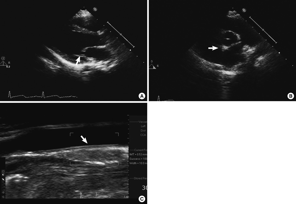
Fig. 2 (A) Echocardiographic parasternal long-axis view showes mitral annulus and mitral valve calcification compatible with senile process. (B) Echocardiographic parasternal short-axis view of arotic valve showes inhomogeneously increased echodensity of the valve and thickening of the leaflets. (C) Echocardiographic view of left commom carotid artery shows that the wall is thicker than the mean chronological age's.
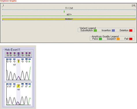
Fig. 3 Sequence analysis of exon 11 of LMNA gene. This result shows G608G (GGC->GGT) heterogenous substitution in LMNA gene.
Reference
-
1. Rastogi R, Chander Mohan SM. Progeria syndrome: a case report. Indian J Orthop. 2008. 42:97–99.2. Gordon LB, Brown WT, Collins FS. Pagon RA, Bird TD, Dolan CR, Stephens K, editors. Hutchinson-Gilford Progeria Syndrome. GeneReviews [Internet]. 1993-. Seattle: University of Washington;2003 Dec 12 [updated 2011 Jan 06].3. Agarwal US, Sitaraman S, Mehta S, Panse G. Hutchinson-Gilford progeria syndrome. Indian J Dermatol Venereol Leprol. 2010. 76:591.4. Cho MH, Choi YW, Kim WS, Lee OK, Lee MH. A case of Hutchinson Gilford progeria syndrome. J Korean Pediatr Soc. 1986. 29:106–110.5. Khang SO, Lee JH, Seol IJ, Choi GJ, Lee KS. A case of Hutchinson-Gilford progeria syndrome. J Korean Pediatr Soc. 1985. 28:405–410.6. Jeoung BJ, Kim DH. A case of Hutchinson-Gilford progeria syndrome. J Korean Pediatr Soc. 1986. 29:90–94.7. Lee MW, Lee BG, Hwang PH, Lee DY, Kim JS. Hutchinson-Gilford progeria syndrome. J Korean Pediatr Soc. 1992. 35:971–977.8. Ha JW, Shim WH, Yoon JH, Chung NS, Lee WK. Cardiovascular findings of Hutchinson-Gilford progeria syndrome. Korean J Intern Med. 1991. 41:572–577.9. Ha JW, Shim WH, Chung NS. Cardiovascular findings of Hutchinson-Gilford syndrome: a Doppler and two-dimensional echocardiographic study. Yonsei Med J. 1993. 34:352–355.10. Gordon LB, McCarten KM, Giobbie-Hurder A, Machan JT, Campbell SE, Berns SD, Kieran MW. Disease progression in Hutchinson-Gilford progeria syndrome: impact on growth and development. Pediatrics. 2007. 120:824–833.11. Eriksson M, Brown WT, Gordon LB, Glynn MW, Singer J, Scott L, Erdos MR, Robbins CM, Moses TY, Berglund P, Dutra A, Pak E, Dukin S, Csoka AB, Boehnke M, Glover TW, Collins FS. Recurrent de novo point mutation in lamin A cause Hutchinson-Gilford progeria syndrome. Nature. 2003. 423:293–298.12. Kudlow BA, Kennedy BK, Monnat RJ Jr. Werner and Hutchinson-Gilford progeria syndromes: mechanistic basis of human progeroid diseases. Nat Rev Mol Cell Biol. 2007. 8:394–404.13. Sullivan T, Escalante-Alcalde D, Bhatt H, Anver M, Bhat N, Nagashima K, Stewart Cl, Burke B. Loss of A-type lamin expression compromises nuclear envelope integrity leading to muscular dystrophy. J Cell Biol. 1999. 147:913–920.14. Capell BC, Erdos MR, Madigan JP, Fiordalisi JJ, Varga R, Conneely KN, Gordon LB, Der CJ, Cox AD, Collins FS. Inhibiting farnesylation of progerin prevents the characteristic nuclear blebbing of Hutchinson-Gilford progeria syndrome. Proc Natl Acad Sci U S A. 2005. 102:12879–12884.15. Lima LL, Ribas CB, Pereira PM, Schettini RA, Eiras Jda D. Do you know this syndrome? Huntchinson-Gilford syndrome (progeria). An Bras Dermatol. 2011. 86:165–166.16. Doll RJ, Kirschmeier P, Bishop WR. Farnesyltransferase inhibitors as anticancer agents: critical crossroads. Curr Opin Drug Discov Devel. 2004. 7:478–486.17. Bishop WR, Bond R, Petrin J, Wang L, Patton R, Doll R, Njoroge G, Catino J, Schwartz J, Windsor W, Syto R, Schwartz J, Carr D, James L, Kirschmeier P. Novel tricyclic inhibitors of farnesyl protein transferase. Biochemical characterization and inhibition of Ras modification in transfected Cos cells. J Biol Chem. 1995. 270:30611–30618.18. Beck LA, Hosick TJ, Sinensky M. Isoprenylation is required for the processing of the lamin A precursor. J Cell Biol. 1990. 110:1489–1499.19. Pollex RL, Hegele RA. Hutchinson-Gilford progeria syndrome. Clin Genet. 2004. 66:375–381.
- Full Text Links
-
- Actions
-
Cited
- CITED
-
- Close
- Share
-
- Similar articles
-
- Hutchinson-Gilford progeria syndrome
- A Case of Hutchinson-Gilford Progeria Syndrome
- Detection of Cerebrovascular Disease in a Child with Hutchinson-Gilford Progeria Syndrome Using MR Angiography: A Case Report
- A Case of Hutchinson-Gilford Progeria Syndrome
- Cardiovascular findings of Hutchinson-Gilford progeria syndrome
