J Periodontal Implant Sci.
2013 Feb;43(1):18-23. 10.5051/jpis.2013.43.1.18.
Bone apposition on implants coated with calcium phosphate by ion beam assisted deposition in oversized drilled sockets: a histologic and histometric analysis in dogs
- Affiliations
-
- 1Department of Periodontology, Research Institute for Periodontal Regeneration, Yonsei University College of Dentistry, Seoul, Korea. shchoi726@yuhs.ac
- 2Department of Periodontology, Seoul National University School of Dentistry, Seoul, Korea.
- 3Institute of Physics and Applied Physics, Atomic-Scale Surface Science Research Center, Yonsei University, Seoul, Korea.
- KMID: 1783673
- DOI: http://doi.org/10.5051/jpis.2013.43.1.18
Abstract
- PURPOSE
The purpose of this study was to evaluate the osseointegration of calcium phosphate (CaP)-coated implants by ion beam assisted deposition with a lack of primary stability.
METHODS
A total of 20 CaP-coated implants were bilaterally placed in the mandible of five dogs. In the rotational implant group, the implants were inserted in oversized drilled sockets without mechanical engagement, while the conventional surgical protocol was followed in the control group. Each group was allowed to heal for 4 and 8 weeks. The bone-to-implant contact (BIC, %) was measured by a histometric analysis.
RESULTS
All of the implants were well-maintained and healing was uneventful. In the histologic observation, all of the implants tested were successfully osseointegrated with a high level of BIC at both observation intervals. There was no significant difference in BIC among any of the groups.
CONCLUSIONS
Within the limitation of this study, successful osseointegration of CaP-coated implants could be achieved in unfavorable conditions without primary stability.
MeSH Terms
Figure
-
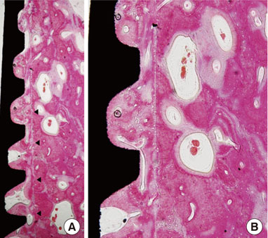
Figure 1 Rotational implants group, 4-week healing period (H&E: A, ×40; B, ×100). Arrowheads: reversal line representing implant ostectomy.
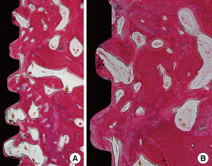
Figure 2 Control group, 4-week healing period (H&E: A, ×40; B, ×100).
Figure 3 Rotational implants group, 8-week healing period (H&E, ×200). Primary and secondary osteons within the thread area. Note that the reversal line still remains.
Figure 4 Control group, 8-week healing period (H&E, ×200). Cell-rich bone marrow zone was seen.
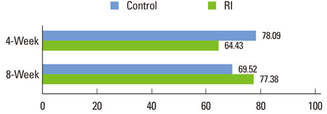
Figure 5 Mean bone-to-implant contact (%) in the six most coronal threads. RI: rotational implants.
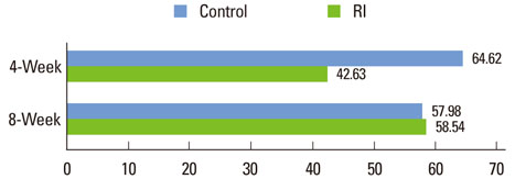
Figure 6 Mean bone density (%) in the six most coronal threads. RI: rotational implants.
Reference
-
1. Albrektsson T, Jansson T, Lekholm U. Osseointegrated dental implants. Dent Clin North Am. 1986. 30:151–174.2. Branemark PI. Osseointegration and its experimental background. J Prosthet Dent. 1983. 50:399–410.
Article3. Cochran DL. A comparison of endosseous dental implant surfaces. J Periodontol. 1999. 70:1523–1539.
Article4. Botticelli D, Berglundh T, Buser D, Lindhe J. The jumping distance revisited: an experimental study in the dog. Clin Oral Implants Res. 2003. 14:35–42.5. Botticelli D, Berglundh T, Lindhe J. Resolution of bone defects of varying dimension and configuration in the marginal portion of the peri-implant bone. An experimental study in the dog. J Clin Periodontol. 2004. 31:309–317.
Article6. Botticelli D, Berglundh T, Persson LG, Lindhe J. Bone regeneration at implants with turned or rough surfaces in self-contained defects: an experimental study in the dog. J Clin Periodontol. 2005. 32:448–455.
Article7. Junker R, Dimakis A, Thoneick M, Jansen JA. Effects of implant surface coatings and composition on bone integration: a systematic review. Clin Oral Implants Res. 2009. 20:Suppl 4. 185–206.
Article8. Shalabi MM, Gortemaker A, Van't Hof MA, Jansen JA, Creugers NH. Implant surface roughness and bone healing: a systematic review. J Dent Res. 2006. 85:496–500.
Article9. Jung UW, Hwang JW, Choi DY, Hu KS, Kwon MK, Choi SH, et al. Surface characteristics of a novel hydroxyapatite-coated dental implant. J Periodontal Implant Sci. 2012. 42:59–63.
Article10. Lacefield WR. Hydroxyapatite coatings. Ann N Y Acad Sci. 1988. 523:72–80.
Article11. Hayashi K, Inadome T, Mashima T, Sugioka Y. Comparison of bone-implant interface shear strength of solid hydroxyapatite and hydroxyapatite-coated titanium implants. J Biomed Mater Res. 1993. 27:557–563.
Article12. van Dijk K, Schaeken HG, Wolke JC, Maree CH, Habraken FH, Verhoeven J, et al. Influence of discharge power level on the properties of hydroxyapatite films deposited on Ti6A14V with RF magnetron sputtering. J Biomed Mater Res. 1995. 29:269–276.
Article13. Lee IS, Zhao B, Lee GH, Choi SH, Chung SM. Industrial application of ion beam assisted deposition on medical implants. Surf Coat Technol. 2007. 201:5132–5137.
Article14. Wikesjo UM, Susin C, Qahash M, Polimeni G, Leknes KN, Shanaman RH, et al. The critical-size supraalveolar peri-implant defect model: characteristics and use. J Clin Periodontol. 2006. 33:846–854.
Article15. Jung UW, Kim CS, Choi SH, Cho KS, Inoue T, Kim CK. Healing of surgically created circumferential gap around non-submerged-type implants in dogs: a histomorphometric study. Clin Oral Implants Res. 2007. 18:171–178.
Article16. Davies JE. Understanding peri-implant endosseous healing. J Dent Educ. 2003. 67:932–949.
Article17. Sivolella S, Bressan E, Salata LA, Urrutia ZA, Lang NP, Botticelli D. Osteogenesis at implants without primary bone contact: an experimental study in dogs. Clin Oral Implants Res. 2012. 23:542–549.
Article18. Araujo MG, Lindhe J. Dimensional ridge alterations following tooth extraction. An experimental study in the dog. J Clin Periodontol. 2005. 32:212–218.
Article19. Lazzara RJ. Immediate implant placement into extraction sites: surgical and restorative advantages. Int J Periodontics Restorative Dent. 1989. 9:332–343.20. Stadlinger B, Pourmand P, Locher MC, Schulz MC. Systematic review of animal models for the study of implant integration, assessing the influence of material, surface and design. J Clin Periodontol. 2012. 39:Suppl 12. 28–36.
Article21. Chae GJ, Jung UW, Jung SM, Lee IS, Cho KS, Kim CK, et al. Healing of surgically created circumferential gap around nano-coating surface dental implants in dogs. Surf Interface Anal. 2008. 40:184–187.
Article22. Jung UW, Kim S, Kim YH, Cha JK, Lee IS, Choi SH. Osseointegration of dental implants installed without mechanical engagement: a histometric analysis in dogs. Clin Oral Implants Res. 2012. 23:1297–1301.
Article23. Song JE, Yoon HJ, Um YJ, Chae GJ, Jung UW, Chung SM, et al. The effect of a multi-treated implant surface on gap defect healing in dogs. Thin Solid Films. 2009. 517:5352–5356.
Article24. Um YJ, Song JE, Chae GJ, Jung UW, Chung SM, Lee IS, et al. The effect of post heat treatment of hydroxyapatite-coated implants on the healing of circumferential coronal defects in dogs. Thin Solid Films. 2009. 517:5375–5379.
Article
- Full Text Links
-
- Actions
-
Cited
- CITED
-
- Close
- Share
-
- Similar articles
-
- Dissolution behavior and early bone apposition of calcium phosphate-coated machined implants
- A STUDY OF BONE APPOSITION AND MARGINAL ALVEOLAR BONE LOSS AROUND IMMEDIATE IMPLANSTS
- The histometric analysis of osseointegration in hydroxyapatite surface dental implants by ion beam-assisted deposition
- Histomorphometric study of machined titanium implants and calcium phosphate coated titanium implants
- Surface analyses of titanium substrate modified by anodization and nanoscale Ca-P deposition
