Imaging Features of Mucinous Breast Carcinoma
- Affiliations
-
- 1Department of Radiology, College of Medicine, The Catholic University of Korea, Korea. rad-ksh@catholic.ac.kr
- 2Department of Pathology, College of Medicine, The Catholic University of Korea, Korea.
- KMID: 1782990
- DOI: http://doi.org/10.13104/jksmrm.2010.14.1.21
Abstract
- PURPOSE
To examine the imaging findings of mucinous breast carcinoma and to evaluate the difference in these findings based on the histopathologic grade.
MATERIALS AND METHODS
We retrospectively analyzed the imaging features according to BI-RADS in 29 patients with surgically proven mucinous carcinoma. The histopathologic grade was classified as well-differentiated, moderately-differentiated and poorly-differentiated. Based on these criteria, the differences in imaging findings were statistically analyzed.
RESULTS
Mammography was available in 20 cases, which contained 17 mass lesions (85%) and 3 cases of normal findings. On ultrasonography (27 cases), mucinous carcinoma was observed as a mass with an oval shape (59.3%), a microlobulated margin (55.6%) or an inhomogeneous isoechogenicity (74.1%). On MRI (21 cases), mucinous carcinoma was commonly observed to have a lobular shape (76%), smooth margin (86%) or heterogeneous contrast-enhancement (61.9%). On the kinetic curve, there was a delayed wash-out pattern (52.3%). There were no significant differences in the imaging findings for each histopathologic grade except that a well-differentiated tumor had an abrupt interface.
CONCLUSION
A well-differentiated mucinous carcinoma tended to have an abrupt interface on ultrasonography, as compared with the moderately-differentiated one. Mucinous carcinoma showed a heterogeneous enhancement and a delayed washout kinetic curve pattern on dynamic MRI.
Keyword
MeSH Terms
Figure
-

Fig. 1 Left mucinous carcinoma in a 54-year-old woman. (a) Mammogram showed a lobular, circumscribed, isodense mass without calcifications. (b) Ultrasonography showed an oval, microlobulated, heterogeneously isoechoic mass with posterior enhancement and abrupt interface.
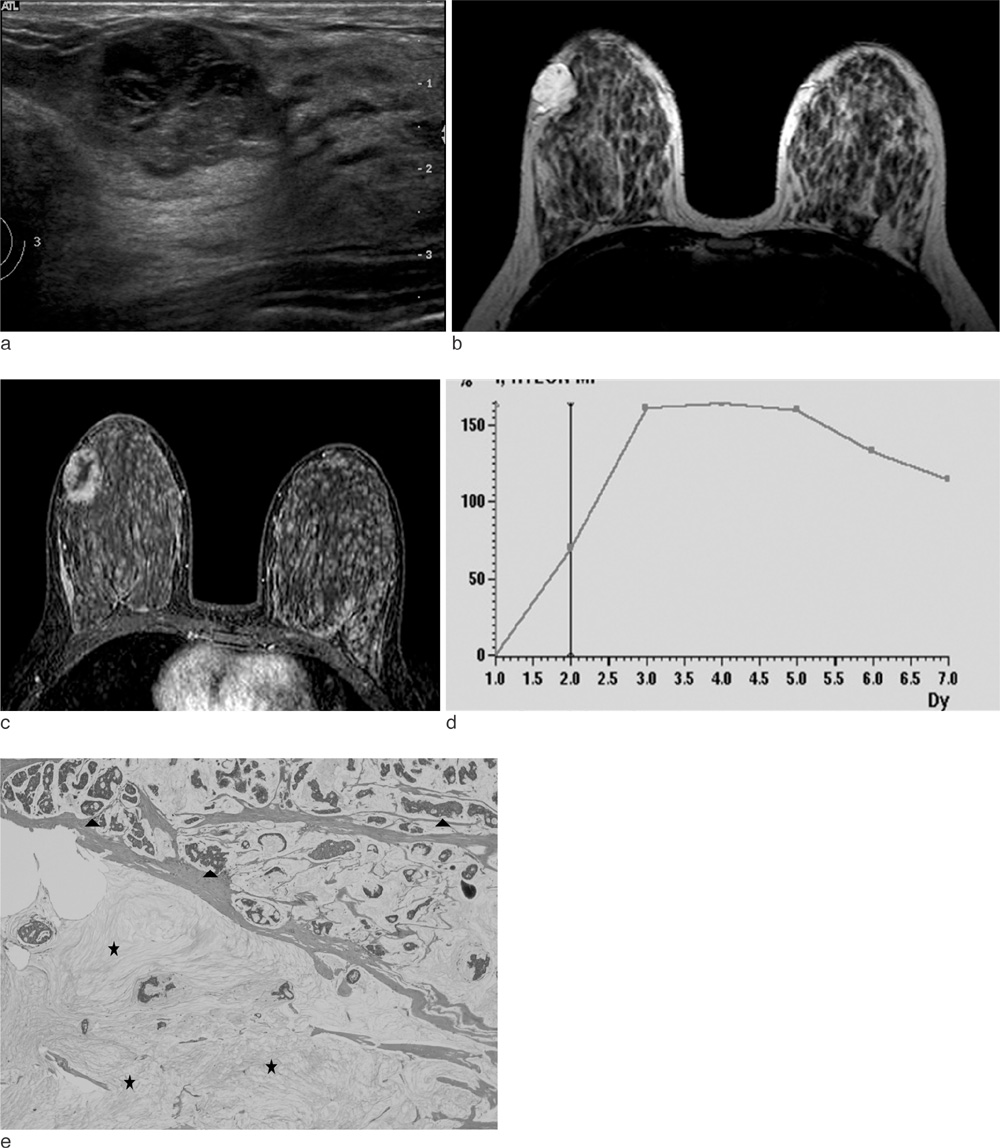
Fig. 2 Right mucinous carcinoma in a 41-year-old woman. (a) Ultrasonography showed an oval, microlobulated, complex echoic mass with posterior enhancement and abrupt interface. (b) Axial T2-weighted image showed a hyperintense, lobular mass with a smooth border. (c) Axial contrast-enhanced T1-weighted image showed rim enhancement. (d) Kinetic curve showed the delayed washout pattern. (e) Mucinous carcinoma consisted of hypocellular areas with large lakes filled with mucin and sparse clusters of tumor cells (black stars), and hypercellular areas with crowded clusters of tumor cells in the mucin (black arrowheads) (Hematoxylin-eosin; magnification ×40).
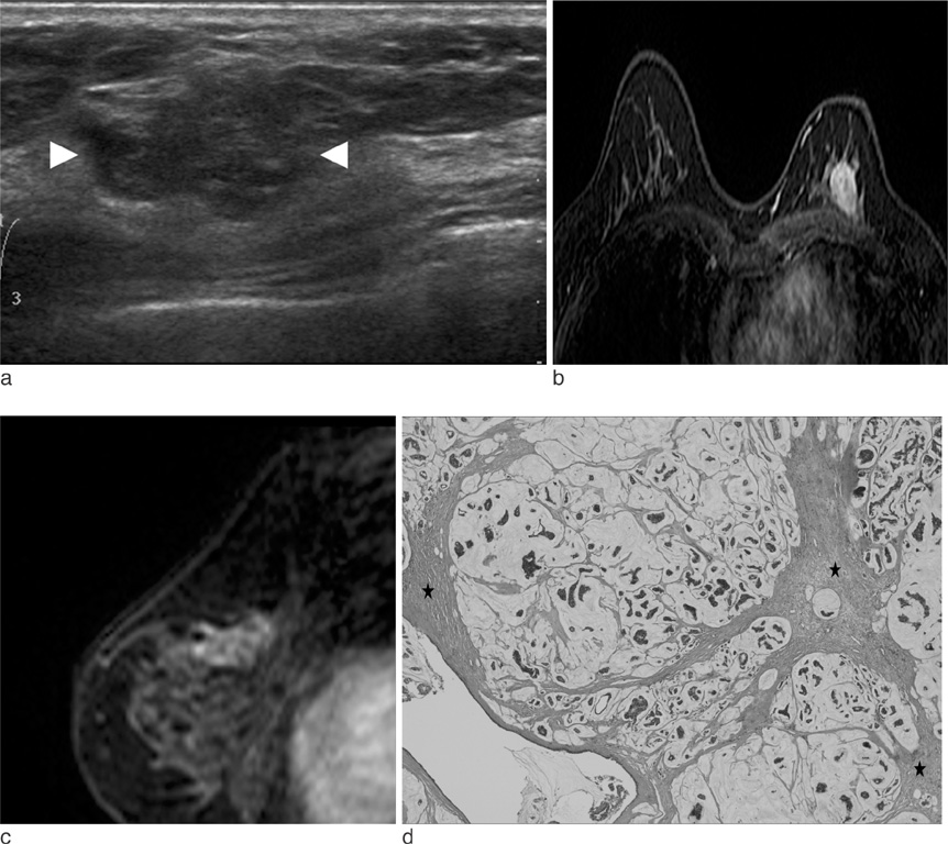
Fig. 3 Left mucinous carcinoma in a 52-year-old woman. (a) Ultrasonography showed an irregular, microlobulated and heterogeneously isoechoic mass with posterior-enhancement and an echogenic halo (white arrowheads). (b, c) Axial and sagittal contrast-enhanced T1-weighted image showed an oval, heterogeneously enhancing mass with irregular margin. (d) Mucin pools of variable sizes were separated by fibrous septae (black stars) and clusters of tumor cells floating in the mucin pools (Hematoxylin-eosin; magnification ×40).
Reference
-
1. Conant EF, Dillon RL, Palazzo J, Ehrlich SM, Feig SA. Imaging findings in mucin-containing carcinomas of the breast: correlation with pathologic features. AJR Am J Roentgenol. 1994. 163:821–824.2. Dhillon R, Depree P, Metcalf C, Wylie E. Screen-detected mucinous carcinoma: potential for delayed diagnosis. Clin Radiol. 2006. 61:423–430.3. Cardenosa G, Doudna C, Eklund GW. Mucinous (colloid) breast cancer: clinical and mammographic findings in 10 patients. AJR Am J Roentgenol. 1994. 162:1077–1079.4. Wilson TE, Helvie MA, Oberman HA, Joynt LK. Pure and mixed mucinous carcinoma of the breast: pathologic basis for differences in mammographic appearance. AJR Am J Roentgenol. 1995. 165:285–289.5. Matsuda M, Yoshimoto M, Iwase T, et al. Mammographic and clinicopathological features of mucinous carcinoma of the breast. Breast Cancer. 2000. 7:65–70.6. Kushwaha AC, Whitman GJ, Williamson JD. Mucinous carcinoma of the breast. AJR Am J Roentgenol. 1999. 173:290.7. Goodman DN, Boutross-Tadross O, Jong RA. Mammographic features of puremucinous carcinoma of the breast with pathological correlation. Can Assoc Radiol J. 1995. 46:296–301.8. Svane G, Potchen EJ, Sierra A, Azavedo E. Screening mammography. 1993. St. Louis, MO: Mosby;296–299. 308–311.9. Kopans DB. Breast imaging. 1989. Philadelphia: Lippincott;298.10. Parsons CA. Diagnosis Of Breast Disease: Imaging, Clinical Features And Pathology. 1983. Baltimore: University Park Press;142–162.11. Pina Insausti LJ, Soga Garcia E. Mucinous breast carcinoma showing as a cluster of suspicious microcalcifications on mammography. Eur Radiol. 1998. 8:1666–1668.12. Memis A, Ozdemir N, Parildar M, Ustunn EE, Erhan Y. Mucinous breast cancer: mammographic and US features with histologic correlation. Eur J Radiol. 2000. 35:39–43.13. Lam WW, Chu WC, Tse GM, Ma TK. Sonographic appearance of mucinous carcinoma of the breast. AJR Am J Roentgenol. 2004. 182:1069–1074.14. Chopra S, Evans AJ, Pinder SE, et al. Pure mucinous breast cancer: mammographic and ultrasound findings. Clin Radiol. 1996. 51:421–424.15. Stavros AT. Stavros AT, editor. Malignant solid breast nodules: specific types. Breast ultrasound. 2003. Philadelphia: Lipincott Williams & Wilkins;645–648.16. Kamitani K, Ono M, Toyoshima S, et al. Isoechoic axillary lymph node metastases of mucinous carcinoma of the breast: a case report. Breast cancer. 2006. 13:382–385.17. Kawashima M, Tamaki Y, Nonaka T, et al. MR imaging of mucinous carcinoma of the breast. AJR Am J Roentgenol. 2002. 179:179–183.18. Okafuji T, Yabuuchi H, Sakai S. MR imaging features of pure mucinous carcinoma of the breast. Eur J Radiol. 2006. 60:405–413.19. Yuen S, Uematsu T, Kasami M, et al. Breast carcinomas with strong high-signal intensity on T2-weighted MR images: pathological characteristics and differential diagnosis. J Magn Reson Imaging. 2007. 25:502–510.20. Monzawa S, Yokokawa M, et al. Mucinous Carcinoma of the Breast: MRI Features of Pure and Mixed Forms with Histopathologic Correlation. AJR Am J Roentgenol. 2009. 192:W125–W131.21. Clayton F. Pure mucinous carcinoma of the breast: morphologic features and prognostic correlates. Hum Pathol. 1986. 17:34–38.22. Wedegartner U, Bick U, Wortler K, Rummery E, Bongartz G. Differentiation between benign and malignant findings on MR-mammography: usefulness of morphological criteria. Eur Radiol. 2001. 11:1645–1650.
- Full Text Links
-
- Actions
-
Cited
- CITED
-
- Close
- Share
-
- Similar articles
-
- Mucinous carcinoma of the breast: distinctive histopathologic and genetic characteristics
- MR Imaging of Mucinous Carcinoma of the Breast Associated with Ductal Carcinoma In Situ: Case Report
- MR Imaging Findings and Texture Analysis of Pure and Mixed Mucinous Breast Carcinoma
- Clinicopathologic Analysis of 40 Mucinous Breast Carcinomas
- Prognostic Significance of a Micropapillary Pattern in Pure Mucinous Carcinoma of the Breast: Comparative Analysis with Micropapillary Carcinoma
