Radiation Exposure of the Hand and Chest during C-arm Fluoroscopy-Guided Procedures
- Affiliations
-
- 1Department of Anesthesiology and Pain Medicine, Konkuk University Medical Center, Seoul, Korea. painfree@kuh.ac.kr
- 2Department of Anesthesiology and Pain Medicine, Seoul National University Hospital, Seoul, Korea.
- KMID: 1779024
- DOI: http://doi.org/10.3344/kjp.2013.26.1.51
Abstract
- BACKGROUND
The C-arm fluoroscope is an essential tool for the intervention of pain. The aim of this study was to investigate the radiation exposure experienced by the hand and chest of pain physicians during C-arm fluoroscopy-guided procedures.
METHODS
This is a prospective study about radiation exposure to physicians during transforaminal epidural steroid injection (TFESI) and medial branch block (MBB). Four pain physicians were involved in this study. Data about effective dose (ED) at each physician's right hand and left side of the chest, exposure time, radiation absorbed dose (RAD), and the distance from the center of the X-ray field to the physician during X-ray scanning were collected.
RESULTS
Three hundred and fifteen cases were included for this study. Demographic data showed no significant differences among the physicians in the TFESIs and MBBs. In the TFESI group, there was a significant difference between the ED at the hand and chest in all the physicians. In physician A, B and C, the ED at the chest was more than the ED at the hand. The distance from the center of the X-ray field to physician A was more than that of the other physicians, and for the exposure time, the ED and RAD in physician A was less than that of the other physicians. In the MBB group, there was no difference in the ED at the hand and chest, except for physician D. The distance from the center of the X-ray field to physician A was more than that of the other physicians and the exposure time in physician A was less than that of the other physicians.
CONCLUSIONS
In conclusion, the distance from the radiation source, position of the hand, experience and technique can correlate with the radiation dose.
Keyword
Figure
-
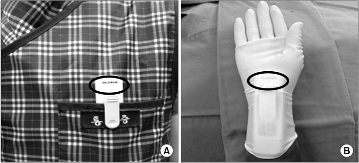
Fig. 1 Two dosimeters were used to record the radiation exposure. (A) One was worn on the left chest over the lead apron. (B) The other was worn on the palmar aspect of the right hand. Circle: detector of dosimeter.

Fig. 2 Various position of hands during X-ray scanning. (A) Physician A's position, (B) Physician B and C's position, (C) Physician D's position.
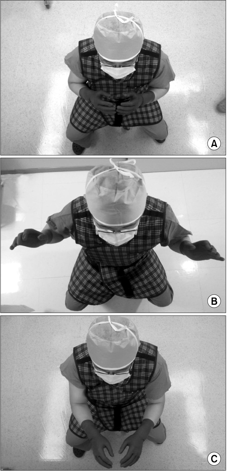
Fig. 3 The positions of each physician's hand which was looked down. (A) Physician A's position, (B) Physician B and C's position, (C) Physician D's position.
Cited by 8 articles
-
Radiation exposure to the eyes and thyroid during C-arm fluoroscopy-guided cervical epidural injections is far below the safety limit
Eun Joo Choi, Gwangcheol Go, Woong Ki Han, Pyung-Bok Lee
Korean J Pain. 2020;33(1):73-80. doi: 10.3344/kjp.2020.33.1.73.Replay to the Letter: Are Doctors Exposed to Radiation Even When Wearing Protectors during Fluoroscopic Procedures?
Jae Hun Kim
Korean J Pain. 2013;26(2):208-208. doi: 10.3344/kjp.2013.26.2.208.Are Doctors Exposed to Radiation Even When Wearing Protectors during Fluoroscopic Procedures?
Dae Hyun Jo
Korean J Pain. 2013;26(2):207-207. doi: 10.3344/kjp.2013.26.2.207.Radiation Safety for Pain Physicians: Technique or Equipment
Jae Hang Shim
Korean J Pain. 2014;27(2):101-102. doi: 10.3344/kjp.2014.27.2.101.How Effective Are Radiation Reducing Gloves in C-arm Fluoroscopy-guided Pain Interventions?
Ah Na Kim, Young Jae Chang, Bo Kyung Cheon, Jae Hun Kim
Korean J Pain. 2014;27(2):145-151. doi: 10.3344/kjp.2014.27.2.145.The Radiation Exposure of Radiographer Related to the Location in C-arm Fluoroscopy-guided Pain Interventions
Young Jae Chang, Ah Na Kim, In Su Oh, Nam Sik Woo, Hae Kyoung Kim, Jae Hun Kim
Korean J Pain. 2014;27(2):162-167. doi: 10.3344/kjp.2014.27.2.162.Three principles for radiation safety: time, distance, and shielding
Jae Hun Kim
Korean J Pain. 2018;31(3):145-146. doi: 10.3344/kjp.2018.31.3.145.Radiation safety for pain physicians: principles and recommendations
Sewon Park, Minjung Kim, Jae Hun Kim
Korean J Pain. 2022;35(2):129-139. doi: 10.3344/kjp.2022.35.2.129.
Reference
-
1. Andreassi MG. The biological effects of diagnostic cardiac imaging on chronically exposed physicians: the importance of being non-ionizing. Cardiovasc Ultrasound. 2004; 2:25. PMID: 15555078.
Article2. Park PE, Park JM, Kang JE, Cho JH, Cho SJ, Kim JH, et al. Radiation safety and education in the applicants of the final test for the expert of pain medicine. Korean J Pain. 2012; 25:16–21. PMID: 22259711.
Article3. Pradhan AS. Evolution of dosimetric quantities of International Commission on Radiological Protection (ICRP): Impact of the forthcoming recommendations. J Med Phys. 2007; 32:89–91. PMID: 21157526.
Article4. Miller DL, Vañó E, Bartal G, Balter S, Dixon R, Padovani R, et al. Occupational radiation protection in interventional radiology: a joint guideline of the Cardiovascular and Interventional Radiology Society of Europe and the Society of Interventional Radiology. J Vasc Interv Radiol. 2010; 21:607–615. PMID: 20430294.
Article5. Harding LK, Thomson WH. International commission on radiation protection. Nucl Med Commun. 1990; 11:585–587. PMID: 2234693.
Article6. Cousins C, Sharp C. Medical interventional procedures-reducing the radiation risks. Clin Radiol. 2004; 59:468–473. PMID: 15145716.
Article7. Mroz TE, Yamashita T, Davros WJ, Lieberman IH. Radiation exposure to the surgeon and the patient during kyphoplasty. J Spinal Disord Tech. 2008; 21:96–100. PMID: 18391712.
Article8. Theocharopoulos N, Perisinakis K, Damilakis J, Papadokostakis G, Hadjipavlou A, Gourtsoyiannis N. Occupational exposure from common fluoroscopic projections used in orthopaedic surgery. J Bone Joint Surg Am. 2003; 85:1698–1703. PMID: 12954827.
Article9. Blattert TR, Fill UA, Kunz E, Panzer W, Weckbach A, Regulla DF. Skill dependence of radiation exposure for the orthopaedic surgeon during interlocking nailing of long-bone shaft fractures: a clinical study. Arch Orthop Trauma Surg. 2004; 124:659–664. PMID: 15365718.
Article10. Bahari S, Morris S, Broe D, Taylor C, Lenehan B, McElwain J. Radiation exposure of the hands and thyroid gland during percutaneous wiring of wrist and hand procedures. Acta Orthop Belg. 2006; 72:194–198. PMID: 16768265.11. Arnstein PM, Richards AM, Putney R. The risk from radiation exposure during operative X-ray screening in hand surgery. J Hand Surg Br. 1994; 19:393–396. PMID: 8077836.
Article12. Siegel JA, Marcus CS, Sparks RB. Calculating the absorbed dose from radioactive patients: the line-source versus point-source model. J Nucl Med. 2002; 43:1241–1244. PMID: 12215565.13. Bushberg JT, Seibert JA, Leidholdt EM, Boone JM. The essential physics of medical imaging. 2011. 3rd ed. Philadelphia: Lippincott Williams & Wilkins;p. 837–902.14. Singer G. Occupational radiation exposure to the surgeon. J Am Acad Orthop Surg. 2005; 13:69–76. PMID: 15712984.
Article15. Cho JH, Kim JY, Kang JE, Park PE, Kim JH, Lim JA, et al. A study to compare the radiation absorbed dose of the C-arm fluoroscopic modes. Korean J Pain. 2011; 24:199–204. PMID: 22220241.
Article16. Kallmes DF, O E, Roy SS, Piccolo RG, Marx WF, Lee JK, et al. Radiation dose to the operator during vertebroplasty: prospective comparison of the use of 1-cc syringes versus an injection device. AJNR Am J Neuroradiol. 2003; 24:1257–1260. PMID: 12812967.
- Full Text Links
-
- Actions
-
Cited
- CITED
-
- Close
- Share
-
- Similar articles
-
- Radiation Exposure to the Hand of a Spinal Interventionalist during Fluoroscopically Guided Procedures
- Radiation exposure and its reduction in the fluoroscopic examination and fluoroscopy-guided interventional radiology
- Radiation safety for pain physicians: principles and recommendations
- Radiation safety: a focus on lead aprons and thyroid shields in interventional pain management
- Radiation exposure and protection for eyes in pain management
