Synchrotron Microangiography of the Rat Heart Using the Langendorff Model
- Affiliations
-
- 1Department of Thoracic and Cardiovascular Surgery, Seoul National University College of Medicine, Children's Hospital, Seoul, Korea. woonghan@snu.ac.kr
- 2Department of Cardiothoracic Surgery, Sejong General Hospital, Sejong Heart Institute, Bucheon, Korea.
- 3Medical Student, University of New South Wales College of Medicine, Sydney, Australia.
- KMID: 1776346
- DOI: http://doi.org/10.4070/kcj.2008.38.9.462
Abstract
- BACKGROUND AND OBJECTIVES
The ability to study microvessels of a beating heart in real time at the level of the capillary is essential for research. However, there are no proven methods currently available to achieve this. The conventional absorption-contrast agents have limitations for studying capillaries. Microangiography with using synchrotron phase-contrast X-ray technology and no contrast agent has recently been reported on. We tried to verify this previous report, and we wanted to visualize the microvessels of a rat heart using air as a contrast agent. MATERIALS AND METHODS: We made the Langendorff apparatus in a hutch of the Pohang Accelerator Laboratory. The images were obtained with a white beam and a monochromatic beam. The visual images were magnified using 3x and 20x optical microscope lenses, and the images were captured with a charge-coupled device camera. RESULTS: We could not duplicate the previously reported findings in which microvessels were visualized without the use of contrast agent. But with using air as a contrast agent, the microvasculature of rat hearts was clearly identified at a spatial resolution of 1.2 microm. Air being absorbed inside a capillary was also observed. Vessels under 10 microm diameter were unable to be visualized with using iodine as a contrast agent. CONCLUSION: Phase contrast imaging already allows spatial resolution of 1 microm, which is enough to inspect capillaries. We were able to obtain images of cardiac capillaries with using air as a contrast agent. Yet air has the fatal limitations in that it causes embolism and ischemia. A more suitable contrast agent or imaging method needs to be developed in order to study the microvessels of a beating heart.
Keyword
MeSH Terms
Figure
-

Fig. 1 The Langendorff apparatus built in a hutch of the Pohang Accelerator Laboratory. A rat heart was continuously perfused with oxygenated (95% O2/ 5% CO2) and warmed (38℃) Krebs solution.
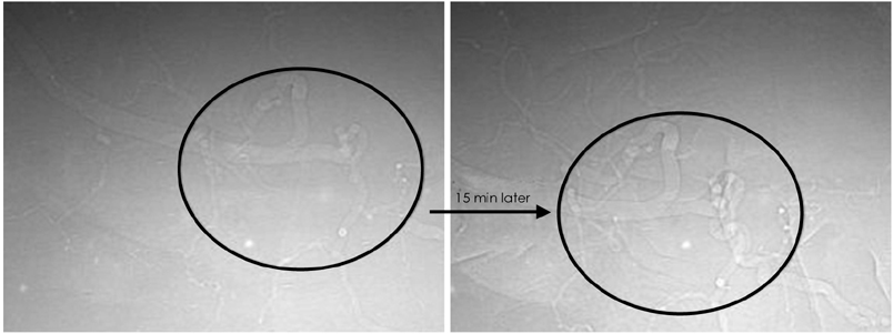
Fig. 2 Vascular torsion and vaporization of the intravascular blood, which were both due to synchrotron radiation heat. The contrast effect became more distinct as time went by.
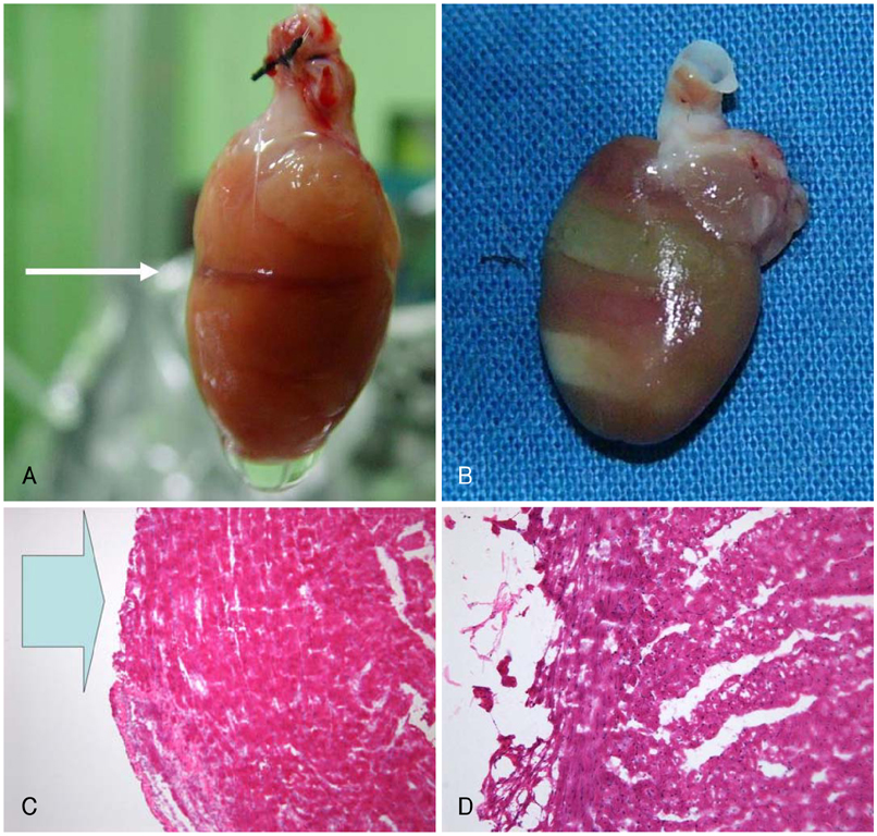
Fig. 3 Gross findings (A, B) and pathologic findings (C, D) of radiation tissue damage using the 7B2 beamline. A: when we exposed the heart to narrow-width radiation, a dark strip of tissue developed. B: we exposed the heart to broad width radiation twice for 15 minutes each time. Two bright strips were obtained. C: pathologic findings (×10) of the specimen and (B), loss of the epicardial tissue was found (large arrow). D: pathologic findings (×20) of the specimen and (B), myocardial damage was observed.

Fig. 4 Coronary angiography with iodine contrast agent in the heart of rat. The major coronary artery and its branch were visualized. However, it was difficult to get an image of the microvasculature.
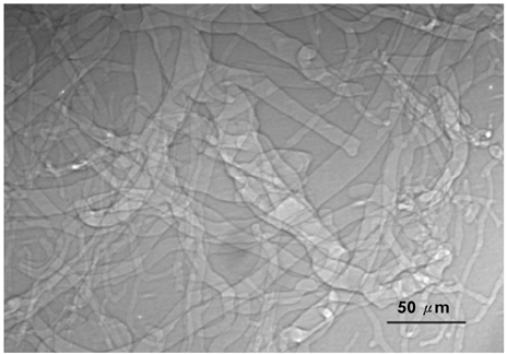
Fig. 5 Microangiography with massive air embolism. We infused massive air (1 cc) into the coronary sinus. We could get the microangiographic images of rat heart at the level of a capillary with using air as a contrast agent.
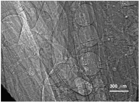
Fig. 6 Phase-contrast synchrotron microangiography of rat toe using air as a contrast agent.
Reference
-
1. Hwu Y, Tsai WL, Groso A, Margaritondo G, Je JH. Coherence-enhanced synchrotron radiology: simple theory and practical applications. J Phys D Appl Phys. 2002. 35:R105–R120.2. Morita M, Ohkawa M, Miyazaki S, Ishimaru T, Umetani K, Suzuki K. Simultaneous observation of superficial cortical and intracerebral microvessels in vivo during reperfusion after transient forebrain ischemia in rats using synchrotron radiation. Brain Res. 2007. 1158:116–122.3. Kidoguchi K, Tamaki M, Mizobe T, et al. In vivo X-ray angiography in the mouse brain using synchrotron radiation. Stroke. 2006. 37:1856–1861.4. Takeshita S, Isshiki T, Ochiai M, et al. Endothelium-dependent relaxation of collateral microvessels after intramuscular gene transfer of vascular endothelial growth factor in a rat model of hindlimb ischemia. Circulation. 1998. 98:1261–1263.5. Takeda T, Momose A, Wu J, et al. Vessel imaging by interferometric phase-contrast X-ray technique. Circulation. 2002. 105:1708–1712.6. Hwu Y, Tsai WL, Je JH, et al. Synchrotron microangiography with no contrast agent. Phys Med Biol. 2004. 49:501–508.7. Kim JW, Seo HS, Hwu Y, et al. In vivo real-time vessel imaging and ex vivo 3D reconstruction of atherosclerotic plaque in apolipoprotein E-knockout mice using synchrotron radiation microscopy. Int J Cardiol. 2007. 114:166–171.8. Baik S, Kim HS, Jeong MH, et al. International consortium on phase contrast imaging and radiology beamline at the Pohang Light Source. Rev Sci Instrum. 2004. 75:4355–4358.9. Mori H, Hyodo K, Tobita K, et al. Visualization of penetrating transmural arteries in situ by monochromatic synchrotron radiation. Circulation. 1994. 89:863–871.10. Yamashita T, Kawashima S, Ozaki M, et al. Mouse coronary angiograph using synchrotron radiation microangiography. Circulation. 2002. 105:E3–E4.11. Takeshita S, Isshiki T, Mori H, et al. Use of synchrotron radiation microangiography to assess development of small collateral arteries in a rat model of hindlimb ischemia. Circulation. 1997. 95:805–808.12. Ohtsuka S, Sugishita Y, Takeda T, et al. Dynamic intravenous coronary angiography using 2D monochromatic synchrotron radiation. Br J Radiol. 1999. 72:24–28.13. Blattmann H, Gebbers JO, Bräuer-Krisch E, et al. Applications of synchrotron X-rays to radiotherapy. Nucl Instrum Methods Phys Res A. 2005. 548:17–22.
- Full Text Links
-
- Actions
-
Cited
- CITED
-
- Close
- Share
-
- Similar articles
-
- Effect of L-carnitine on ischemic myocardium of Langendorff's isolated rat heart
- Technique of Renal Microangiography
- An experimental study on vascular changes in renal biopsy injury
- Effect of High Doses of Propofol on Myocardium in the Isolated Rat Heart
- An experimental study on the normal brain microangiography
