Evaluation of the Significance of Incidental Breast Lesions Detected by Chest CT
- Affiliations
-
- 1Department of Radiology, Soonchunhyang University College of Medicine, Soonchunhyang University Hospital, Seoul, Korea. ywchang@hosp.sch.ac.kr
- 2Department of Radiology, Soonchunhyang University College of Medicine, Soonchunhyang University Cheonan Hospital, Cheonan, Korea.
- 3Department of Radiology, Soonchunhyang University College of Medicine, Soonchunhyang University Bucheon Hospital, Bucheon, Korea.
- 4Department of Radiology, Soonchunhyang University College of Medicine, Soonchunhyang University Gumi Hospital, Gumi, Korea.
- KMID: 1748457
- DOI: http://doi.org/10.3348/jksr.2013.68.3.229
Abstract
- PURPOSE
To evaluate the significance of incidentally detected breast lesions on a chest CT scan.
MATERIALS AND METHODS
Thirty-six incidental breast lesions in 26 patients were detected on a chest CT scan and were correlated with breast sonography, retrospectively. Among them, twenty-four breast lesions in 20 patients that were correlated with chest CT and sonography were available to pathology or follow up sonography. The CT findings were compared with sonographic findings according to the pathologic results.
RESULTS
Incidentally detected breast lesions on a chest CT scan were correlated with sonography in 86% (31/36). Among 24 lesions that were available to pathology or follow up sonography, seven (29.2%) lesions were malignant and 17 (70.8%) lesions were benign. CT revealed a significant difference between benign and malignant lesions in terms of shape and margin (p = 0.007; p = 0.008, respectively). The CT findings were well correlated with sonographic findings in shape and margin (p = 0.001, respectively).
CONCLUSION
Incidentally detected breast lesions on chest CT can be correlated with sonography. An irregular shape or a non-circumscribed margin of breast lesions on a CT scan can be considered as a suggestive sign of malignancy.
Figure
-
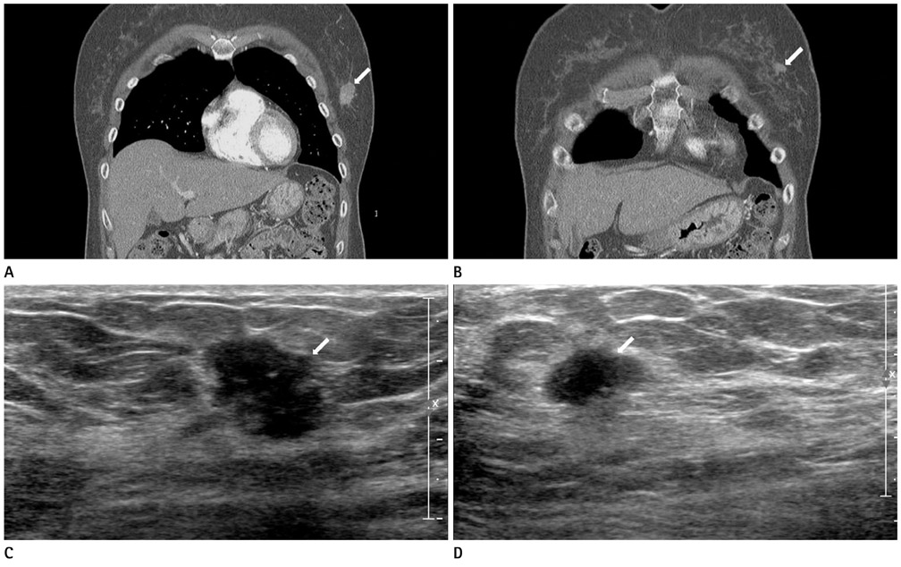
Fig. 1 A 53-year-old woman underwent chest CT after traumatic contusion. A, B. Chest CT with multiplanar reconstruction imaging shows microlobulated, irregular, and inhomogeneous enhancing mass in the left breast [Hounsfield unit (HU) = 127] (arrow). Another mass of the same nature was detected in the left breast (HU = 91) (arrow). C, D. Sonography shows irregular, ill defined, hypoechogenic masses in the left breast (arrow). These masses were confirmed to be malignant on biopsy.
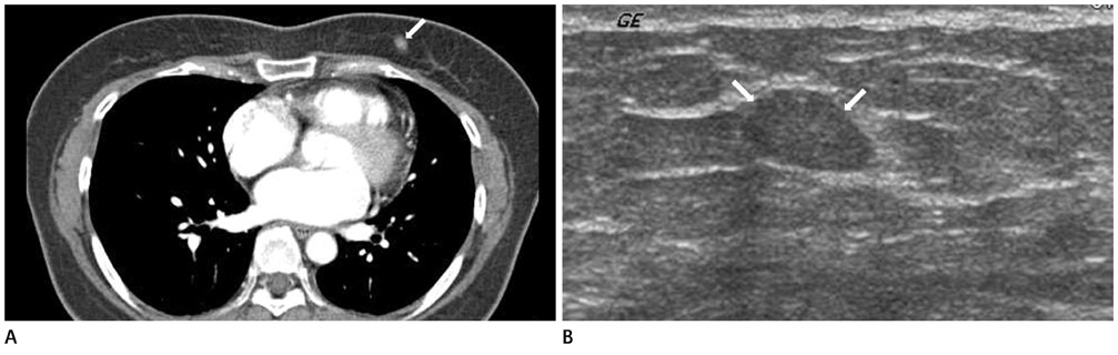
Fig. 2 A 50-year-old women underwent chest CT due to palpitations. A. Chest CT scan shows a well-defined, round, homogeneous enhancing mass in the left breast measuring 7 mm (Hounsfield unit = 99) (arrow). B. Sonography shows a well-circumscribed, oval, isoechogenic mass in the left breast (arrows). The pathological diagnosis was fibroadenoma with fibrocystic change.
Cited by 1 articles
-
Incidental Breast Lesions on Chest CT: Clinical Significance and Differential Features Requiring Referral
Yun Jung Choi, Tae Hoon Kim, Yoon Jin Cha, Eun Ju Son, Hye Mi Gweon, Chul Hwan Park
J Korean Soc Radiol. 2018;79(6):303-310. doi: 10.3348/jksr.2018.79.6.303.
Reference
-
1. Harish MG, Konda SD, MacMahon H, Newstead GM. Breast lesions incidentally detected with CT: what the general radiologist needs to know. Radiographics. 2007. 27:Suppl 1. S37–S51.2. Yi JG, Kim SJ, Marom EM, Park JH, Jung SI, Lee MW. Chest CT of incidental breast lesions. J Thorac Imaging. 2008. 23:148–155.3. Porter G, Steel J, Paisley K, Watkins R, Holgate C. Incidental breast masses detected by computed tomography: are any imaging features predictive of malignancy? Clin Radiol. 2009. 64:529–533.4. Lin WC, Hsu HH, Li CS, Yu JC, Hsu GC, Yu CP, et al. Incidentally detected enhancing breast lesions on chest computed tomography. Korean J Radiol. 2011. 12:44–51.5. Inoue M, Sano T, Watai R, Ashikaga R, Ueda K, Watatani M, et al. Dynamic multidetector CT of breast tumors: diagnostic features and comparison with conventional techniques. AJR Am J Roentgenol. 2003. 181:679–686.6. Kang DK, Kim MJ, Jung YS, Yim H. Clinical application of multidetector row computed tomography in patient with breast cancer. J Comput Assist Tomogr. 2008. 32:583–598.7. Hussain A, Gordon-Dixon A, Almusawy H, Sinha P, Desai A. The incidence and outcome of incidental breast lesions detected by computed tomography. Ann R Coll Surg Engl. 2010. 92:124–126.8. Moyle P, Sonoda L, Britton P, Sinnatamby R. Incidental breast lesions detected on CT: what is their significance? Br J Radiol. 2010. 83:233–240.9. Kim SM, Park JM. Computed tomography of the breast. Abnormal findings with mammographic and sonographic correlation. J Comput Assist Tomogr. 2003. 27:761–777.10. Prionas ND, Lindfors KK, Ray S, Huang SY, Beckett LA, Monsky WL, et al. Contrast-enhanced dedicated breast CT: initial clinical experience. Radiology. 2010. 256:714–723.
- Full Text Links
-
- Actions
-
Cited
- CITED
-
- Close
- Share
-
- Similar articles
-
- Incidentally Detected Enhancing Breast Lesions on Chest Computed Tomography
- Incidental Breast Lesions on Chest CT: Clinical Significance and Differential Features Requiring Referral
- Incidental Musculoskeletal Lesions Detected on Abdominopelvic CT Scans: A Pictorial Essay
- Detection of Breast Abnormalities on Enhanced Chest CT: Correlation with Breast Composition on Mammography
- The Evaluation of Contralateral Breast Lesions in Breast Cancer Patients Using Reduction Mammoplasty
