Incidental Breast Lesions on Chest CT: Clinical Significance and Differential Features Requiring Referral
- Affiliations
-
- 1Department of Radiology, Gangnam Severance Hospital, Yonsei University College of Medicine, Seoul, Korea. park-chulhwan@yuhs.ac
- 2Department of Pathology, Gangnam Severance Hospital, Yonsei University College of Medicine, Seoul, Korea.
- KMID: 2427377
- DOI: http://doi.org/10.3348/jksr.2018.79.6.303
Abstract
- PURPOSE
To evaluate the CT features of incidental breast lesions on chest CT and to suggest useful criteria for referral to a specialized breast unit.
MATERIALS AND METHODS
Between May 2009 and April 2014, enhanced chest CT examination reports containing the key word "˜breast' were reviewed retrospectively. Patients who had incidental breast lesion and were referred to a specialized breast unit and then underwent pathological confirmation or follow-up over a 1-year period were included. Finally, 86 patients (all female, mean age, 48.9 ± 12.6 years) were enrolled. Two radiologists evaluated lesion characteristics, including size, shape, margins, and enhancement. The correlations between the CT features and pathologies were evaluated, and the diagnostic accuracy of CT features in various combinations was assessed.
RESULTS
Among the CT features, irregular shape, non-circumscribed margin, and high contrast enhancement were different between malignant and benign lesions (p < 0.05). The combination of non-circumscribed margin and high contrast enhancement had the highest accuracy (97.7%).
CONCLUSION
Reliable CT features for incidental malignant breast masses are irregular shape, non-circumscribed margin, and high contrast enhancement. The combination of non-circumscribed margin and high contrast enhancement could help distinguish incidental malignant breast lesions and indicate referral to a specialized breast unit.
MeSH Terms
Figure
-
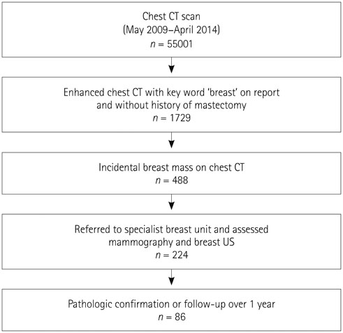
Fig. 1 Flow chart of patient selection.
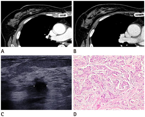
Fig. 2 A 60-year-old woman underwent chest CT for a medical examination. A. Axial CT reveals a round, non-circumscribed mass with high contrast enhancement in the right upper medial quadrant of the breast. B. Two years later, tumor size increased from 8 mm to 10 mm. C. Sonography of the right breast at the corresponding location reveals hypoechoic mass with irregular margin. D. Pathological examination revealed infiltrative ductal carcinoma with desmoplastic stroma (hematoxylin-eosin stain, × 200).
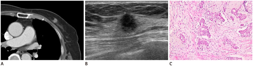
Fig. 3 An 86-year-old woman underwent CT for a thyroid mass. A. Axial CT scan reveals a non-circumscribed, irregular mass with high contrast enhancement in the left upper medial quadrant of the breast. B. Sonography reveals a hypoechoic mass with irregular margin. C. Pathological examination revealed infiltrative ductal carcinoma (hematoxylin-eosin stain, × 200).
Reference
-
1. Skinner S. Guide to thoracic imaging. Aust Fam Physician. 2015; 44:558–562.2. Yi JG, Kim SJ, Marom EM, Park JH, Jung SI, Lee MW. Chest CT of incidental breast lesions. J Thorac Imaging. 2008; 23:148–155.
Article3. Monzawa S, Washio T, Yasuoka R, Mitsuo M, Kadotani Y, Hanioka K. Incidental detection of clinically unexpected breast lesions by computed tomography. Acta Radiol. 2013; 54:374–379.
Article4. Cho EM, Kang H, Shin YG, Yun JH, Oh KS, Park S. Detection of breast abnormalities on enhanced chest CT: correlation with breast composition on mammography. J Korean Soc Radiol. 2017; 76:96–103.
Article5. Kim JH, Chang YW, Hwang JH, Kim HH, Lee EH, Yang SB. Evaluation of the significance of incidental breast lesions detected by chest CT. J Korean Soc Radiol. 2013; 68:229–235.
Article6. Goldberg PA, White CS, McAvoy MA, Templeton PA. CT appearance of the normal and abnormal breast with mammographic correlation. Clin Imaging. 1994; 18:262–272.7. Jacobs PC, Mali WP, Grobbee DE, van der Graaf Y. Prevalence of incidental findings in computed tomographic screening of the chest: a systematic review. J Comput Assist Tomogr. 2008; 32:214–221.8. Shojaku H, Seto H, Iwai H, Kitazawa S, Fukushima W, Saito K. Detection of incidental breast tumors by noncontrast spiral computed tomography of the chest. Radiat Med. 2008; 26:362–367.
Article9. Hussain A, Gordon-Dixon A, Almusawy H, Sinha P, Desai A. The incidence and outcome of incidental breast lesions detected by computed tomography. Ann R Coll Surg Engl. 2010; 92:124–126.
Article10. Porter G, Steel J, Paisley K, Watkins R, Holgate C. Incidental breast masses detected by computed tomography: are any imaging features predictive of malignancy? Clin Radiol. 2009; 64:529–533.
Article11. Bach AG, Abbas J, Jasaabuu C, Schramm D, Wienke A, Surov A. Comparison between incidental malignant and benign breast lesions detected by computed tomography: a systematic review. J Med Imaging Radiat Oncol. 2013; 57:529–533.
Article12. Lin WC, Hsu HH, Li CS, Yu JC, Hsu GC, Yu CP, et al. Incidentally detected enhancing breast lesions on chest computed tomography. Korean J Radiol. 2011; 12:44–51.
Article13. Harish MG, Konda SD, MacMahon H, Newstead GM. Breast lesions incidentally detected with CT: what the general radiologist needs to know. Radiographics. 2007; 27:Suppl 1. S37–S51.
Article14. Jakanani G, Al-Attar M. RE: incidental breast masses detected by computed tomography: are any imaging features predictive of malignancy? Clin Radiol. 2009; 64:1041–1042.
Article15. Prionas ND, Lindfors KK, Ray S, Huang SY, Beckett LA, Monsky WL, et al. Contrast-enhanced dedicated breast CT: initial clinical experience. Radiology. 2010; 256:714–723.
Article16. Inoue M, Sano T, Watai R, Ashikaga R, Ueda K, Watatani M, et al. Dynamic multidetector CT of breast tumors: diagnostic features and comparison with conventional techniques. AJR Am J Roentgenol. 2003; 181:679–686.17. Litmanovich D, Gourevich K, Israel O, Gallimidi Z. Unexpected foci of 18F-FDG uptake in the breast detected by PET/CT: incidence and clinical significance. Eur J Nucl Med Mol Imaging. 2009; 36:1558–1564.18. D'Orsi CJ. ACR BI-RADS Atlas: breast imaging reporting and data system. 5th ed. Reston, VA: American College of Radiology;2013.19. Mendelson E, Böhm-Vélez M, Berg W, Whitman G, Feldman M, Madjar H. ACR BI-RADS® ultrasound. In : D'Orsi CJ, editor. ACR BI-RADS Atlas: breast imaging reporting and data system. 5th ed. Reston, VA: American College of Radiology;2013. p. 35–100.20. Sickles E, D'Orsi C, Bassett L, Appleton C, Berg W, Burnside E. ACR BI-RADS® mammography. In : D'Orsi CJ, editor. ACR BI-RADS® Atlas, breast imaging reporting and data system. Reston, VA: American College of Radiology;2013. 5:p. 13–117.21. Meller MT, Cox JE, Callanan KW. Incidental detection of breast lesions with computed tomography. Clin Breast Cancer. 2007; 7:634–637.
Article22. Uematsu T, Sano M, Homma K. False-positive helical CT findings of multifocal and multicentric breast cancer: is attenuation of tumor useful for diagnosing enhanced lesions? Breast Cancer. 2002; 9:62–68.
Article23. Sardanelli F, Calabrese M, Zandrino F, Melani E, Parodi R, Imperiale A, et al. Dynamic helical CT of breast tumors. J Comput Assist Tomogr. 1998; 22:398–407.
Article24. Kim SM, Park JM. Computed tomography of the breast. Abnormal findings with mammographic and sonographic correlation. J Comput Assist Tomogr. 2003; 27:761–777.
- Full Text Links
-
- Actions
-
Cited
- CITED
-
- Close
- Share
-
- Similar articles
-
- Evaluation of the Significance of Incidental Breast Lesions Detected by Chest CT
- Incidentally Detected Enhancing Breast Lesions on Chest Computed Tomography
- Detection of Breast Abnormalities on Enhanced Chest CT: Correlation with Breast Composition on Mammography
- Incidental Musculoskeletal Lesions Detected on Abdominopelvic CT Scans: A Pictorial Essay
- Incidental Extramammary Findings on Preoperative Breast MRI in Breast Cancer Patients: A Pictorial Essay
