Astroblastoma: A Case Report
- Affiliations
-
- 1Department of Pathology, Yeungnam University College of Medicine, Daegu, Korea.
- 2Department of Pathology, Catholic University of Daegu School of Medicine, Daegu, Korea. dskim@yumail.ac.kr
- 3Department of Neurosurgery, Catholic University of Daegu School of Medicine, Daegu, Korea.
- KMID: 1733527
- DOI: http://doi.org/10.3346/jkms.2004.19.5.772
Abstract
- Astroblastoma is one of the very unusual type of tumors, whose histogenesis has not been clarified. It occurs mainly among children or young adults. Astroblastoma is grossly well-demarcated, and shows histologically characteristic perivascular pseudorosettes with frequent vascular hyalinization. Perivascular pseudorosettes in astroblastoma have short and thick cytoplasmic processes and blunt-ended foot plates. A 15-yr-old girl presented with headache and diplopia for one and a half year. A welldemarcated mass, 9.7 cm in diameter, was found in the right frontal lobe in brain MRI, and it was a well-enhanced inhomogenous mass. Cystic changes of various sizes were observed inside the tumor mass as well as in the posterior part of the mass, but no peritumoral edema was found. Histologically, this mass belongs to a typical astroblastoma, and no sign of anaplastic astrocytoma, gemistocytic astrocytoma or glioblastoma was found in any part of the tumor. Immunohistochemically, the tumor cells showed diffuse strong positivity for glial fibrillary acidic protein, S-100 protein, vimentin and neuron specific enolase, and focal positivity for epithelial membrane antigen and CAM 5.2, while showing negativity for synaptophysin, neurofilament protein, pan-cytokeratin and high molecular weight keratin.
Keyword
MeSH Terms
-
Adolescent
Brain Neoplasms/metabolism/*pathology
Diagnosis, Differential
Female
Glial Fibrillary Acidic Protein/metabolism
Humans
Keratin/metabolism
Magnetic Resonance Imaging
Neoplasms, Neuroepithelial/metabolism/*pathology
Phosphopyruvate Hydratase/metabolism
Research Support, Non-U.S. Gov't
S100 Proteins/metabolism
Vimentin/metabolism
Figure
-
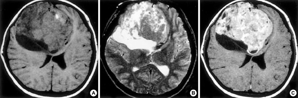
Fig. 1 (A) T1-weighted image shows a huge well-demarcated mass in the right frontal lobe. (B) Many cystic changes of different sizes are observed within the tumor on T2-weighted image. (C) The tumor shows an inhomogenous enhancement.
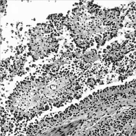
Fig. 2 Well-developed perivascular pseudorosettes are seen throughout the tumor (H&E, ×40).
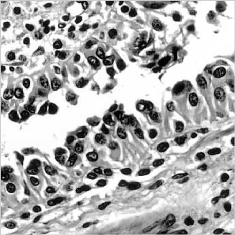
Fig. 3 The tumor cells composing perivascular pseudorosettes display short and thick cytoplasmic processes with prominent blunt-ended footplates toward the vessel wall (H&E, ×400).
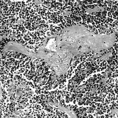
Fig. 4 The fibrovascular stroma frequently shows prominent vascular sclerosis (H&E, ×40).
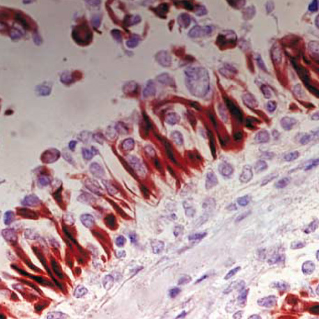
Fig. 5 The broad GFAP-positive glial processes surround blood vessels (×400).

Fig. 6 The tumor cells are positive for S-100 protein (A), vimentin (B), and CAM 5.2 (C) (×200).
Reference
-
1. Brat DJ, Hirose Y, Cohen KJ, Feuerstein BG, Burger PC. Astroblastoma: Clinicopathologic features and chromosomal abnormalities defined by comparative genomic hybridization. Brain Pathol. 2000. 10:342–352.
Article2. Pizer BL, Moss T, Oakhill A, Webb D, Coakham HB. Congenital astroblastoma: an immunohistochemical study. Case report. J Neurosurg. 1995. 83:550–555.3. Lantos PL, Rosenblum MK. Kleihues P, Cavenee WK, editors. Astroblastoma. Pathology and genetics of tumors of the nervous system. 2000. 2nd ed. Lyon: International Agency for Research on Cancer;88–89.4. Bailey P, Bucy PC. Astroblastomas of the brain. Acta Psychiatr Neurol. 1930. 439–461.5. Yamashita J, Handa H, Yamagami T, Haebara H. Astroblastoma of pure type. Surg Neurol. 1985. 24:218–222.
Article6. Hoag G, Sima AA, Rozdilsky B. Astroblastoma revisited: a report of three cases. Acta Neuropathol (Berl). 1986. 70:10–16.
Article7. Husain AN, Leestma JE. Cerebral astroblastoma: immunohistochemical and ultrastructural features. Case report. J Neurosurg. 1986. 64:657–661.8. Bonnin JM, Rubinstein LJ. Astroblastomas: A pathological study of 23 tumors, with a postoperative follow-up in 13 patients. Neursurgery. 1989. 25:6–13.
Article9. Thiessen B, Finlay J, Kulkarni R, Rosenblum MK. Astroblastoma: Does histology predict biologic behavior? J Neurooncol. 1998. 40:59–65.10. Burger PC, Scheithauer BW. Tumors of the central nervous system. Atlas of tumor Pathology. 1994. 3rd series. Washington DC: Armed Forces Institute of Pathology;146–148.11. McLendon RE, Enterline DS, Tien RD, Thorstad WL, Bruner JM. Bigner DD, McLendon RE, Bruner JM, editors. Astroblastomas. Russell & Rubinstein's pathology of tumors of the nervous system. 1998. 6th ed. London: Arnold;419–426.12. Cabello A, Madero S, Castresana A, Diaz-Lobato R. Astroblastoma: Electron microscopy and immunohistochemical findings: Case report. Surg Neurol. 1991. 35:116–121.
Article13. Jay V, Edwards V, Squire J, Rutka J. Astroblastoma: Report of a case with ultrastructural, cell kinetic, and cytogenetic analysis. Pediatr Pathol. 1993. 13:323–332.
Article14. Brat DJ, Cohen KJ, Sanders JM, Geuerstein BG, Burger PC. Clinicopathologic features of astroblastoma.[Abstract]. J Neuropathol Exp Neurol. 1999. 58:509.15. Rubinstein LJ, Herman MM. The astroblastoma and its possible cytogenetic relationship to the tanycyte. Acta Neuropathol. 1989. 78:472–483.16. Lantos PL, Vendenberg SR, Kleihues P. Graham DI, Lantos PL, editors. Tumors of the nervous system. Greenfield's neuropathology. 1997. 6th ed. London: Arnold;647–652.17. Shim YR, Gu MJ, Kim DS, Kim OL, Byun WM, Kim YJ. Choroid plexus carcinoma. A report of two cases. Korean J Pathol. 2001. 35:176–179.
- Full Text Links
-
- Actions
-
Cited
- CITED
-
- Close
- Share
-
- Similar articles
-
- A Case of Astroblastoma
- A Cerebral Astroblastoma Mimicking an Extra-axial Neoplasm
- Astroblastoma in a Young Female Patient: A Case Report and Literature Review of Clinicopathological, Radiological and Prognostic Characteristics and Current Treatment Strategies
- Melorheostosis: A Case Report
- Amelia of Both Lower Extremities: A Case Report
