Primary Central Nervous System ALK Positive Anaplastic Large Cell Lymphoma with Predominantly Leptomeningeal Involvement in an Adult
- Affiliations
-
- 1Department of Neurosurgery, Samsung Medical Center, Sungkyunkwan University School of Medicine, Seoul, Korea.
- 2Department of Pathology, Samsung Medical Center, Sungkyunkwan University School of Medicine, Seoul, Korea. yhko310@skku.edu
- 3Department of Internal Medicine, Samsung Medical Center, Sungkyunkwan University School of Medicine, Seoul, Korea.
- 4Department of Neurosurgery, Konkuk University Chungju Hospital, Chungju, Korea.
- 5Department of Pathology, Ewha Womans University School of Medicine, Seoul, Korea.
- KMID: 1727900
- DOI: http://doi.org/10.3349/ymj.2013.54.3.791
Abstract
- A 31-year-old Korean male presented with altered consciousness and severe headache. Brain MRI delineated focal leptomeningeal enhancement without any intracerebral lesions. Diagnosis was made based on a brain biopsy showing anaplastic large cell lymphoma (ALCL), immunohistochemical stains revealing positivity for anaplastic lymphoma kinase (ALK) and an absence of involvement in any other organs; specifically, the primary central nervous system ALK+ALCL. Complete remission was achieved following 5 cycles of systemic chemotherapy with a high dose of Methotrexate and a simultaneous 7 cycles of intrathecal triple chemotherapy. Diagnosis of primary leptomeningeal ALK+ALCL is challenging given its rarity and non-specific symptoms along with non-pathognomonic radiologic findings. We present the first case of primary leptomeningeal ALK-positive ALCL where the clinical course, pathologic characteristics and treatment modality are described as well as a review of literature.
MeSH Terms
-
Adult
Antineoplastic Agents/therapeutic use
Biopsy
Brain/metabolism/pathology
Diagnosis, Differential
Humans
Immunohistochemistry
Lymphoma, Large-Cell, Anaplastic/*diagnosis/drug therapy/pathology
Male
Meningeal Neoplasms/*diagnosis/drug therapy/pathology
Receptor Protein-Tyrosine Kinases/*metabolism
Antineoplastic Agents
Receptor Protein-Tyrosine Kinases
Figure
-
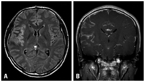
Fig. 1 Magnetic resonance imaging of his brain showed hyperintense signal in the right temporal sulci from the Flair image (A) and leptomeningeal enhancement in the right temporal and insular gyri from Gadolinium enhanced image (B).

Fig. 2 The histologic section showed brain parenchyma infiltrated by numerous small-to-medium sized neoplastic cells with perivascular cuffing of neoplastic cells (A, HE ×100) and the neoplastic cells had irregular nuclei with a moderate amount of cytoplasm (B, HE ×400). Large atypical cells with horseshoe shaped nuclei, which are hallmark cells of anaplastic large cell lymphoma, were also present (B, inset, HE ×1000)
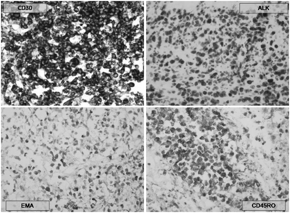
Fig. 3 Immunohistochemical stains revealed immunopositivity for CD30, ALK, Granzyme B, CD45RO and EMA. ALK, anaplastic lymphoma kinase.

Fig. 4 Fluorescence in situ hybridization for translocations involving ALK at 2p23: a unique sequence break apart probe targeting ALK gene locus was used. Translocation involving ALK at 2p23 was detected. ALK, anaplastic lymphoma kinase.
Reference
-
1. Stein H, Mason DY, Gerdes J, O'Connor N, Wainscoat J, Pallesen G, et al. The expression of the Hodgkin's disease associated antigen Ki-1 in reactive and neoplastic lymphoid tissue: evidence that Reed-Sternberg cells and histiocytic malignancies are derived from activated lymphoid cells. Blood. 1985. 66:848–858.2. Jaffe ES, Harris NL, Stein H, Wardiman JW. Tumors of haematopoietic and lymphoid tissues. 2001. Lyon: IARC Press.3. Campo E, Swerdlow SH, Harris NL, Pileri S, Stein H, Jaffe ES. The 2008 WHO classification of lymphoid neoplasms and beyond: evolving concepts and practical applications. Blood. 2011. 117:5019–5032.
Article4. Swerdlow SH, Campo E, Harris NL, Jaffe ES, Pileri SA, Stein H. WHO Classification of Tumours of Haematopoietic and Lymphoid Tissues. 2008. 4th ed. Lyon, France: IARC Press.5. Karikari IO, Thomas KK, Lagoo A, Cummings TJ, George TM. Primary cerebral ALK-1-positive anaplastic large cell lymphoma in a child. Case report and literature review. Pediatr Neurosurg. 2007. 43:516–521.
Article6. Penny RJ, Blaustein JC, Longtine JA, Pinkus GS. Ki-1-positive large cell lymphomas, a heterogenous group of neoplasms. Morphologic, immunophenotypic, genotypic, and clinical features of 24 cases. Cancer. 1991. 68:362–373.
Article7. Falini B, Pileri S, Zinzani PL, Carbone A, Zagonel V, Wolf-Peeters C, et al. ALK+ lymphoma: clinico-pathological findings and outcome. Blood. 1999. 93:2697–2706.8. Abdulkader I, Cameselle-Teijeiro J, Fraga M, Rodriguez-Núnez A, Allut AG, Forteza J. Primary anaplastic large cell lymphoma of the central nervous system. Hum Pathol. 1999. 30:978–981.
Article9. George DH, Scheithauer BW, Aker FV, Kurtin PJ, Burger PC, Cameselle-Teijeiro J, et al. Primary anaplastic large cell lymphoma of the central nervous system: prognostic effect of ALK-1 expression. Am J Surg Pathol. 2003. 27:487–493.10. Ponzoni M, Terreni MR, Ciceri F, Ferreri AJ, Gerevini S, Anzalone N, et al. Primary brain CD30+ ALK1+ anaplastic large cell lymphoma ('ALKoma'): the first case with a combination of 'not common' variants. Ann Oncol. 2002. 13:1827–1832.
Article11. Merlin E, Chabrier S, Verkarre V, Cramer E, Delabesse E, Stéphan JL. Primary leptomeningeal ALK+ lymphoma in a 13-year-old child. J Pediatr Hematol Oncol. 2008. 30:963–967.
Article12. Ozkaynak MF. Favorable outcome of primary CNS anaplastic large cell lymphoma in an immunocompetent patient. J Pediatr Hematol Oncol. 2009. 31:128–130.
Article13. Shiota M, Nakamura S, Ichinohasama R, Abe M, Akagi T, Takeshita M, et al. Anaplastic large cell lymphomas expressing the novel chimeric protein p80NPM/ALK: a distinct clinicopathologic entity. Blood. 1995. 86:1954–1960.
Article14. Villano JL, Koshy M, Shaikh H, Dolecek TA, McCarthy BJ. Age, gender, and racial differences in incidence and survival in primary CNS lymphoma. Br J Cancer. 2011. 105:1414–1418.
Article15. Buxton N, Punt J, Hewitt M. Primary Ki-1-positive T-cell lymphoma of the brain in a child. Pediatr Neurosurg. 1998. 29:250–252.
Article16. Goldbrunner R, Warmuth-Metz M, Tonn JC, Vince GH, Roosen K. Primary Ki-1-positive T-cell lymphoma of the brain--an aggressive subtype of lymphoma: case report and review of the literature. Surg Neurol. 1996. 46:37–41.
Article17. Paulus W, Ott MM, Strik H, Keil V, Müller-Hermelink HK. Large cell anaplastic (KI-1) brain lymphoma of T-cell genotype. Hum Pathol. 1994. 25:1253–1256.
Article18. Lachance DH, O'Neill BP, Macdonald DR, Jaeckle KA, Witzig TE, Li CY, et al. Primary leptomeningeal lymphoma: report of 9 cases, diagnosis with immunocytochemical analysis, and review of the literature. Neurology. 1991. 41:95–100.
Article19. Taga T, Sakaue Y, Anzai Y, Takeuchi Y, Ohta S. Pediatric primary leptomeningeal lymphoma treated without cranial radiotherapy. Pediatr Blood Cancer. 2007. 48:477–478.
Article20. Felice MS, Zubizarreta PA, Rossi JG, Rose A, Alfaro EM, Sackmann-Muriel F. Diagnosis and successful treatment of childhood primary leptomeningeal lymphoma. Med Pediatr Oncol. 2000. 34:361–363.
Article21. Shah AC, Kelly DR, Nabors LB, Oakes WJ, Hilliard LM, Reddy AT. Treatment of primary CNS lymphoma with high-dose methotrexate in immunocompetent pediatric patients. Pediatr Blood Cancer. 2010. 55:1227–1230.
Article
- Full Text Links
-
- Actions
-
Cited
- CITED
-
- Close
- Share
-
- Similar articles
-
- A Case of Multiple Cranial Neuropathies Caused by Anaplastic Lymphoma Kinase-Negative Anaplastic Large Cell Lymphoma
- CD30-Positive Anaplastic Lymphoma Kinase-Negative Systemic Anaplastic Large-Cell Lymphoma in a 9-Year-Old Boy
- A Case of ALK-Negative Systemic Anaplastic Large Cell Lymphoma
- A Case of CD30 (+)/ALK (-) Primary Systemic AnaplasticLarge Cell Lymphoma with Atypical Clinical Features
- A Case of CD30 Positive ALK-Negative Systemic Anaplastic Large Cell Lymphoma Involving Bone Marrow
