Invasive Micropapillary Carcinoma of the Breast: MR Imaging Findings
- Affiliations
-
- 1Department of Radiology, Chonnam National University Medical School, Chonnam National University Hwasun Hospital, Hwasun 519-763, Korea.
- 2Department of Radiology, University of North Carolina at Chapel Hill, NC 27599-7510, USA. cherie_kuzmiak@med.unc.edu
- 3Department of Pathology, Chonnam National University Medical School, Chonnam National University Hwasun Hospital, Hwasun 519-763, Korea.
- 4Department of Surgery, Chonnam National University Medical School, Chonnam National University Hwasun Hospital, Hwasun 519-763, Korea.
- KMID: 1715758
- DOI: http://doi.org/10.3348/kjr.2013.14.4.551
Abstract
OBJECTIVE
To analyze the magnetic resonance (MR) imaging findings of invasive micropapillary carcinoma of the breast.
MATERIALS AND METHODS
MR images were retrospectively evaluated in 14 patients (age range: 37-67, mean age: 49 years) with pathologically confirmed invasive micropapillary carcinoma of the breast. The enhancement type (mass/non-mass), shape, margin, contrast enhancement, and time-intensity curve pattern on the dynamic study were correlated with the histopathologic features. Associated findings, such as edema, nipple change, skin change and enlarged axillary lymph nodes were also studied.
RESULTS
The most common features of the masses were irregular shape (12 of 14 patients, 85.8%) and irregular or spiculated margin (11 of 14 patients, 78.7%). The contrast enhancement was heterogeneous in 11 patients (78.7%), rim enhancement in 2 cases (14.2%), and homogeneous in one patient (7.1%). The predominant kinetic pattern was rapid increase (14 of 14, 100%) in the initial phase and washout (11 of 14, 78.7%) in the delayed phase. Associated non-mass like enhancement was shown in 4 patients, representing ductal carcinoma in situ. MR imaging helped detect additional sites of cancer other than the index lesion in 3 patients (21.4%). Enlarged axillary lymphadenopathy was identified in 7 of the 14 patients (50%).
CONCLUSION
Invasive micropapillary carcinoma appears as a mass with an irregular shape, irregular or spiculated margin and heterogeneous enhancement on MR imaging. Though these findings are not specific and are also observed with other breast malignancies, invasive micropapillary carcinoma frequently showed multiple lesions, accompanying non-mass enhancement and axillary lymph node enlargement.
MeSH Terms
Figure
-
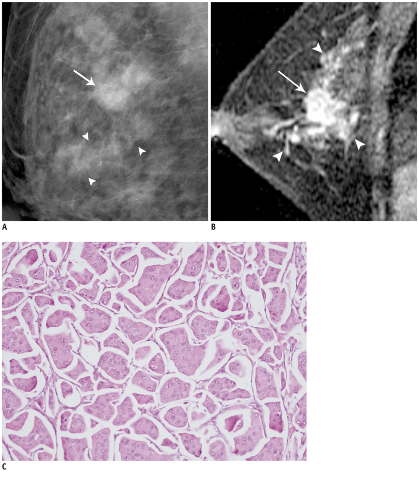
Fig. 1 Forty six-year-old woman with palpable mass in upper outer quadrant of right breast. A. Mediolateral oblique mammogram shows irregular mass (arrow) and segmental distributed pleomorphic microcalcifications (arrowheads) outside of mass, extending anterior to mass. B. Early phase of dynamic enhancement MR imaging shows irregular, heterogeneously enhancing mass (arrow) with adjacent multiple regions of clumped enhancement (arrowheads) extending less than 4.0 cm, representing pathologically proven ductal carcinoma in situ. C. Photomicrograph of histopathological specimen (original magnification, × 100; hematoxylin-eosin stain) of tumor showed small cell clusters surrounded by empty spaces lined by delicate strands of stroma.
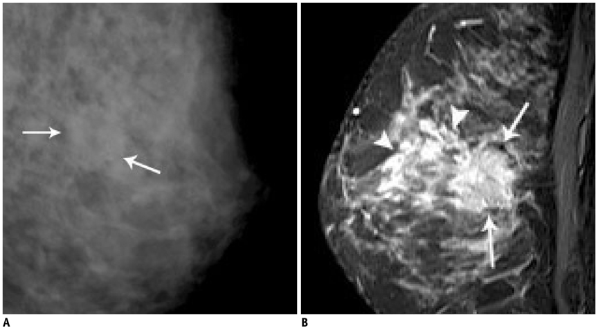
Fig. 2 Thirty nine-year-old woman with palpable mass in left breast. A. Mediolateral oblique mammogram shows irregular equal density mass in left breast corresponding to area of palpation (arrows). B. Dynamic enhancement MR imaging shows irregular heterogeneously enhancing mass (arrows) with segmental clumped enhancement (arrowheads) extending 4 cm anterior to known invasive cancer. MR imaging found additional site. Mastectomy revealed invasive micropapillary carcinoma with extensive ductal carcinoma in situ.
Reference
-
1. Luna-Moré S, Gonzalez B, Acedo C, Rodrigo I, Luna C. Invasive micropapillary carcinoma of the breast. A new special type of invasive mammary carcinoma. Pathol Res Pract. 1994; 190:668–674.2. Paterakos M, Watkin WG, Edgerton SM, Moore DH 2nd, Thor AD. Invasive micropapillary carcinoma of the breast: a prognostic study. Hum Pathol. 1999; 30:1459–1463.3. Kuroda H, Sakamoto G, Ohnisi K, Itoyama S. Clinical and pathologic features of invasive micropapillary carcinoma. Breast Cancer. 2004; 11:169–174.4. Pettinato G, Manivel CJ, Panico L, Sparano L, Petrella G. Invasive micropapillary carcinoma of the breast: clinicopathologic study of 62 cases of a poorly recognized variant with highly aggressive behavior. Am J Clin Pathol. 2004; 121:857–866.5. Yu JI, Choi DH, Park W, Huh SJ, Cho EY, Lim YH, et al. Differences in prognostic factors and patterns of failure between invasive micropapillary carcinoma and invasive ductal carcinoma of the breast: matched case-control study. Breast. 2010; 19:231–237.6. American College of Radiology. Mammography. Breast imaging reporting and data system (BI-RADS). 4th ed. Reston, VA: American College of Radiology;2003.7. American College of Radiology. Ultrasound. Breast imaging reporting and data system (BI-RADS). 4th ed. Reston, VA: American College of Radiology;2003.8. American College of Radiology. MRI. Breast imaging reporting and data system (BI-RADS). 4th ed. Reston, VA: American College of Radiology;2003.9. Liberman L, Morris EA, Dershaw DD, Abramson AF, Tan LK. MR imaging of the ipsilateral breast in women with percutaneously proven breast cancer. AJR Am J Roentgenol. 2003; 180:901–910.10. Siriaunkgul S, Tavassoli FA. Invasive micropapillary carcinoma of the breast. Mod Pathol. 1993; 6:660–662.11. Günhan-Bilgen I, Zekioglu O, Ustün EE, Memis A, Erhan Y. Invasive micropapillary carcinoma of the breast: clinical, mammographic, and sonographic findings with histopathologic correlation. AJR Am J Roentgenol. 2002; 179:927–931.12. Adrada B, Arribas E, Gilcrease M, Yang WT. Invasive micropapillary carcinoma of the breast: mammographic, sonographic, and MRI features. AJR Am J Roentgenol. 2009; 193:W58–W63.13. Kubota K, Ogawa Y, Nishioka A, Murata Y, Itoh S, Hamada N, et al. Radiological imaging features of invasive micropapillary carcinoma of the breast and axillary lymph nodes. Oncol Rep. 2008; 20:1143–1147.14. Ota D, Toyama T, Ichihara S, Mizutani M, Kamei K, Iwata H. A case of invasive micropapillary carcinoma of the breast. Breast Cancer. 2007; 14:323–326.15. Walsh MM, Bleiweiss IJ. Invasive micropapillary carcinoma of the breast: eighty cases of an underrecognized entity. Hum Pathol. 2001; 32:583–589.16. Luna-Moré S, Casquero S, Pérez-Mellado A, Rius F, Weill B, Gornemann I. Importance of estrogen receptors for the behavior of invasive micropapillary carcinoma of the breast. Review of 68 cases with follow-up of 54. Pathol Res Pract. 2000; 196:35–39.17. Nassar H, Wallis T, Andea A, Dey J, Adsay V, Visscher D. Clinicopathologic analysis of invasive micropapillary differentiation in breast carcinoma. Mod Pathol. 2001; 14:836–841.18. Guo X, Chen L, Lang R, Fan Y, Zhang X, Fu L. Invasive micropapillary carcinoma of the breast: association of pathologic features with lymph node metastasis. Am J Clin Pathol. 2006; 126:740–746.
- Full Text Links
-
- Actions
-
Cited
- CITED
-
- Close
- Share
-
- Similar articles
-
- Invasive Micropapillary Carcinoma of the Breast: Mammographic, Sonographic and MR Imaging Findings
- Invasive Micropapillary Carcinoma in Breast Presented as Hyperechoic Mass with Coarse Macrocalcifications: A Case Report
- Invasive Micropapillary Carcinoma of the Breast: A clinicopathologic study of 16 cases
- Fine Needle Aspiration Cytology of Invasive Micropapillary Carcinoma of the Breast
- Prognostic Significance of a Micropapillary Pattern in Pure Mucinous Carcinoma of the Breast: Comparative Analysis with Micropapillary Carcinoma
