A Purely Cystic Giant Sacral Schwannoma Mimicking a Bone Cyst: A Case Report
- Affiliations
-
- 1Department of Radiology, Wonkwang University School of Medicine, Iksan, Korea. medicalq@hanmail.net
- 2Department of Pathology, Wonkwang University School of Medicine, Iksan, Korea.
- KMID: 1709013
- DOI: http://doi.org/10.3348/jksr.2014.71.1.1
Abstract
- Schwannomas are benign tumors arising from the myelinated nerve sheath. These tumors are generally solid, and degenerative changes can occur. However, cystic degeneration is usually noted only in parts of the tumor, and complete cystic change is rare; only a few such cases have been reported. To our knowledge, only one case of a completely cystic giant schwannoma in the lumbosacral area has been reported. Here, we report a similar case with the descriptions of the magnetic resonance imaging and pathological findings.
Figure
-
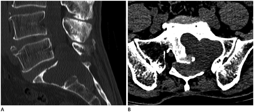
Fig. 1 Lumbar spine computed tomography imaging. A. Sagittal image shows a well-defined osteolytic mass at the sacral region (L5-S2). B. Axial image shows that the mass had a lobulated margin and extended to the intraosseous area of S1 and the epidural space.

Fig. 2 Lumbar spine magnetic resonance imaging. A, B. Sagittal T2-weighted images show a well-defined, unilocular cystic mass. This mass had a fluid-fluid level (or a fluid-debris level) (arrow) and extended to the presacral area (arrowheads). C. Axial T2-weighted image shows that this mass was a purely cystic mass. D. Sagittal T1-weighted image shows the fluid-fluid level (arrow). E. Fat-suppressed contrast enhanced T1-weighted sagittal image shows thin rim enhancement of the cystic mass.
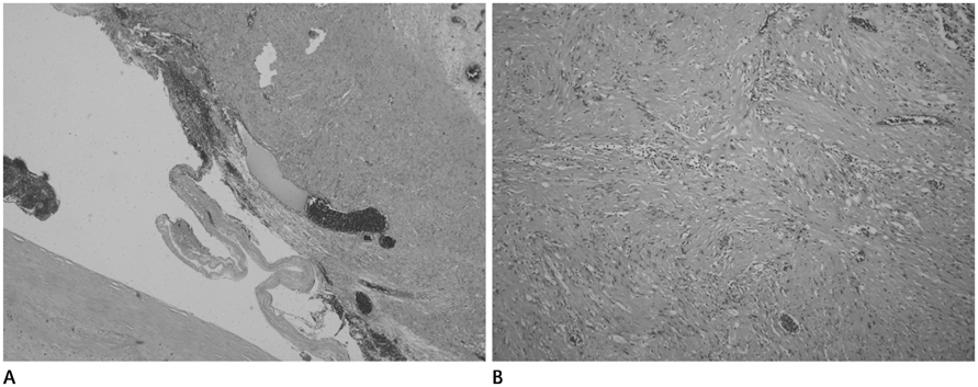
Fig. 3 Microphotography. A. Microphotography (H&E stain, × 40) shows a predominantly cystic component with extensive degeneration and hemorrhage. B. Microphotography (H&E stain, × 100) presents two distinct types of tissue patterns: Antoni A, where the spindle cells are arranged in a relatively compact palisading pattern, and Antoni B, comprising loose, reticular, often cystic arrangements.
Reference
-
1. Abdelwahab IF, Hermann G, Stollman A, Wolfe D, Lewis M, Zawin J. Case report 564: Giant intraosseous schwannoma. Skeletal Radiol. 1989; 18:466–469.2. Conti P, Pansini G, Mouchaty H, Capuano C, Conti R. Spinal neurinomas: retrospective analysis and long-term outcome of 179 consecutively operated cases and review of the literature. Surg Neurol. 2004; 61:34–43. discussion 44.3. Ogose A, Hotta T, Sato S, Takano R, Higuchi T. Presacral schwannoma with purely cystic form. Spine (Phila Pa 1976). 2001; 26:1817–1819.4. Borges G, Bonilha L, Proa M Jr, Fernandes YB, Ramina R, Zanardi V, et al. Imaging features and treatment of an intradural lumbar cystic schwannoma. Arq Neuropsiquiatr. 2005; 63:681–684.5. Nyapathy V, Murthy UK, Chintamani J, Sridhar DY. A case report of a giant presacral cystic schwannoma with sigmoid megacolon. J Radiol Case Rep. 2009; 3:31–37.6. Albert AF, Kirkman MA, du Plessis D, Sacho R, Cowie R, Tzerakis NG. Giant solitary cystic schwannoma of the cervical spine: a case report. Clin Neurol Neurosurg. 2012; 114:396–398.7. Pongsthorn C, Ozawa H, Aizawa T, Kusakabe T, Nakamura T, Itoi E. Giant sacral schwannoma: a report of six cases. Ups J Med Sci. 2010; 115:146–152.8. Wu D, Ba Z, Huang Y, Zhao W, Shen B, Kan H. Totally cystic schwannoma of the lumbar spine. Orthopedics. 2013; 36:e679–e682.9. Sridhar K, Ramamurthi R, Vasudevan MC, Ramamurthi B. Giant invasive spinal schwannomas: definition and surgical management. J Neurosurg. 2001; 94:2 Suppl. 210–215.10. Yu NH, Lee SE, Jahng TA, Chung CK. Giant invasive spinal schwannoma: its clinical features and surgical management. Neurosurgery. 2012; 71:58–67.
- Full Text Links
-
- Actions
-
Cited
- CITED
-
- Close
- Share
-
- Similar articles
-
- Cystic Giant Sacral Schwannoma Mimicking Aneurysmal Bone Cyst : A Case Report and Review of Literatures
- Giant Presacral Schwannoma: A Case Report
- Rare Form of Schwannoma as a Purely Hemorrhagic Cystic Tumor Located in an Intermuscular Plane
- Tibial Schwannoma Mimicking a Popliteal Cyst
- Giant Benign Schwannoma Involving Sacral Bone
