Rare Form of Schwannoma as a Purely Hemorrhagic Cystic Tumor Located in an Intermuscular Plane
- Affiliations
-
- 1Department of Radiology, Inje University Seoul Paik Hospital, College of Medicine, Inje University, Seoul, Korea. jcshim96@unitel.co.kr
- 2Department of Orthopedic Surgery, Inje University Seoul Paik Hospital, College of Medicine, Inje University, Seoul, Korea.
- 3Department of Pathology, Inje University Seoul Paik Hospital, College of Medicine, Inje University, Seoul, Korea.
- KMID: 2376268
- DOI: http://doi.org/10.13104/imri.2017.21.1.38
Abstract
- Schwannomas are mostly solid tumors, some of which may contain cystic degenerations or hemorrhages. However, a schwannoma seen as a purely hemorrhagic cystic tumor is very rare. A 63-year-old woman was referred to the hospital due to a slow-growing mass (present for about 5 years) on her right thigh. She complained about vague pain but without neurologic symptoms such as numbness or tingling sensations. MR images showed an oval lesion with defined margins surrounded by the rectus femoris, vastus lateral, and the vastus intermedius. It was characterized as a multilocular cystic lesion composed of hemorrhagic fluid. In addition, the benign hemorrhagic cystic lesion was differentially diagnosed by radiological techniques as a hemorrhagic ganglion cyst. The lesion was surgically excised and, based on pathological features, was diagnosed as being a schwannoma. We report a purely hemorrhagic cystic schwannoma located in an intermuscular plane.
MeSH Terms
Figure
-

Fig. 1 Schwannoma in a 63-year-old woman. (a) An axial modified proton density image (TR/TE = 2500/33) shows an oval cystic lesion with fluid-fluid level (black arrow). The lesion is surrounded by rectus femoris (*, star shape), vastus lateralis (black arrowhead), and vastus intermedius (white arrowhead) muscles. (b) A sagittal modified proton density image (TR/TE = 2500/33) shows an oval multilocular cystic lesion with low-signal margin. The lesion is surrounded by a rim of fat (split-fat sign) (white arrowheads). (c) Fat-suppressed contrast enhanced T1-weighted axial image (TR/TE = 989/18) shows thin rim enhancement of the cystic mass.
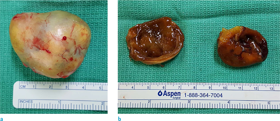
Fig. 2 Photograph of surgical resection specimen of a schwannoma. (a) On gross finding, the lesion is an oval shape and the capsule is yellow-tinged grayish color. (b) Reddish hematoma is noted in the cystic lesion and the surface of the lumen is brown.

Fig. 3 Photomicrograph of a histologic specimen of schwannoma. (a) Pathologic specimen (original magnification, × 100; Hematoxylin-Eosin stain) shows the Antoni A region (not seen in this image) intermingled with the Antoni B region (white arrowhead) in a schwannoma. Wavy, tightly organized nuclear palisades known as Verocay bodies (black arrowheads) occupy the center of a highly cellular field. (b) Pathologic specimen (original magnification, × 200; S-100 stain) shows the universally S-100 stained cells of a schwannoma.
Reference
-
1. Murphey MD, Smith WS, Smith SE, Kransdorf MJ, Temple HT. From the archives of the AFIP. Imaging of musculoskeletal neurogenic tumors: radiologic-pathologic correlation. Radiographics. 1999; 19:1253–1280.2. Ogose A, Hotta T, Koda H, Umezu H, Higuchi T. Images in rheumatology. Hemorrhagic schwannoma with purely cystic appearance in the shoulder. J Rheumatol. 2001; 28:2558–2559.3. Bermejo A, De Bustamante TD, Martinez A, Carrera R, Zabia E, Manjon P. MR imaging in the evaluation of cystic-appearing soft-tissue masses of the extremities. Radiographics. 2013; 33:833–855.4. Wu D, Ba Z, Huang Y, Zhao W, Shen B, Kan H. Totally cystic schwannoma of the lumbar spine. Orthopedics. 2013; 36:e679–e682.5. Lee HA, Jeon SJ, Choi SS, Kim HW, Kim HS. A purely cystic giant sacral schwannoma mimicking a bone cyst: a case report. J Korean Soc Radiol. 2014; 71:1–5.6. Lin J, Martel W. Cross-sectional imaging of peripheral nerve sheath tumors: characteristic signs on CT, MR imaging, and sonography. AJR Am J Roentgenol. 2001; 176:75–82.7. Kwon BC, Baek GH, Chung MS, Lee SH, Kim HS, Oh JH. Intramuscular neurilemoma. J Bone Joint Surg Br. 2003; 85:723–725.8. Shimose S, Sugita T, Kubo T, et al. Major-nerve schwannomas versus intramuscular schwannomas. Acta Radiol. 2007; 48:672–677.9. Knight DM, Birch R, Pringle J. Benign solitary schwannomas: a review of 234 cases. J Bone Joint Surg Br. 2007; 89:382–387.
- Full Text Links
-
- Actions
-
Cited
- CITED
-
- Close
- Share
-
- Similar articles
-
- A Purely Cystic Giant Sacral Schwannoma Mimicking a Bone Cyst: A Case Report
- A case of cystic change of pelvic retroperitoneal Schwannoma misdiagnosed as an ovarian tumor
- Cystic Schwannoma of the Pancreas: A Case Report
- A Case of Purely Epithelioid Peripheral Nerve Sheath Tumor
- Cystic Giant Sacral Schwannoma Mimicking Aneurysmal Bone Cyst : A Case Report and Review of Literatures
