Reversible Verbal and Visual Memory Deficits after Left Retrosplenial Infarction
- Affiliations
-
- 1Department of Neurology, Samsung Medical Center, Sungkyunkwan University School of Medicine, Seoul, Korea. kimgm@smc.samsung.co.kr
- 2Department of Neurology, Chung-Ang University Medical Center, Chung-Ang University School of Medicine, Seoul, Korea.
- KMID: 1700690
- DOI: http://doi.org/10.3988/jcn.2007.3.1.62
Abstract
- The retrosplenial cortex is a cytoarchitecturally distinct brain structure located in the posterior cingulate gyrus and bordering the splenium, precuneus, and calcarine fissure. Functional imaging suggests that the retrosplenium is involved in memory, visuospatial processing, proprioception, and emotion. We report on a patient who developed reversible verbal and visual memory deficits following a stroke. Neuropsychological testing revealed both anterograde and retrograde memory deficits in verbal and visual modalities. Brain diffusion-weighted and T2-weighted magnetic resonance imaging (MRI) demonstrated an acute infarction of the left retrosplenium.
MeSH Terms
Figure
-
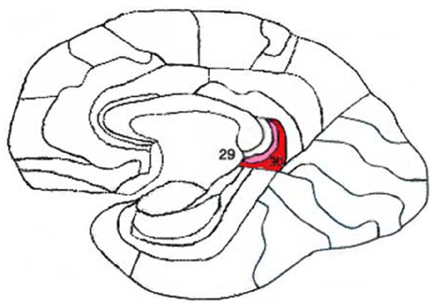
Figure 1 Location of the retrosplenium (Brodmann areas 29 and 30) in a schematic of a brain sagittal section.
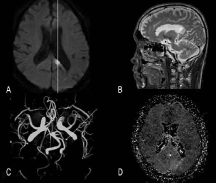
Figure 2 Axial diffusion-weighted magnetic resonance imaging (MRI) demonstrated an acute infarction in the left splenium and retrosplenium. A. White vertical line indicates the plane of the sagittal T2-weighted MRI imaging in panel B. B. sagittal T2-weighted MRI demonstrates that the infarction is restricted to the splenium and retrosplenium. C. Occlusion in the left posterior cerebral artery (PCA) is marked by an arrow in magnetic resonance angiography (MRA). Time-to-peak (TTP) perfusion map reveals a mild perfusion delay to the left occipital region.
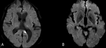
Figure 3 Other lesions were evident in the center of the thalamus (A), and small scattered lesions were evident in the area supplied by the left PCA (B).
Reference
-
1. Saito K, Kimura K, Minematsu K, Shiraishi A, Nakajima M. Transient global amnesia associated with an acute infarction in the retrosplenium of the corpus callosum. J Neurol Sci. 2003. 210:95–97.
Article2. Auchus AP, Chen CP, Sodagar SN, Thong M, Sng EC. Single stroke dementia: insights from 12 cases in Singapore. J Neurol Sci. 2002. 203-204:85–89.3. Valenstein E, Bowers D, Verfaellie M, Heilman KM, Day A, Watson RT. Retrosplenial amnesia. Brain. 1987. 110:1631–1646.
Article4. Maguire EA. The retrosplenial contribution to human navigation: a review of lesion and neuroimaging findings. Scand J Psychol. 2001. 42:225–238.
Article5. Yasuda Y, Watanabe T, Tanaka H, Tadashi I, Akiguchi I. Amnesia following infarction in the right retrosplenial region. Clin Neurol Neurosurg. 1997. 99:102–105.
Article6. Takahashi N, Kawamura M, Shiota J, Kasahata N, Hirayama K. Pure topographic disorientation due to right retrosplenial lesion. Neurology. 1997. 49:464–469.
Article7. Fletcher PC, Frith CD, Grasby PM, Shallice T, Frackowiak RS, Dolan RJ. Brain systems for encoding and retrieval of auditory-verbal memory. An in vivo study in humans. Brain. 1995. 118:401–416.
Article8. Osawa A, Maeshima S, Kubo K, Itakura T. Neuropsychological deficits associated with a tumour in the posterior corpus callosum: a report of two cases. Brain Inj. 2006. 20:673–676.
Article9. Van Groen T, Wyss JM. Connections of the retrosplenial granular b cortex in the rat. J Comp Neurol. 2003. 463:249–263.
Article
- Full Text Links
-
- Actions
-
Cited
- CITED
-
- Close
- Share
-
- Similar articles
-
- The Lateralization and Localization of Memory and Neurocognitive Functioning in Patients with Temporal Lobe Epilepsy
- The Neurocognitive Function Between the Patients Who had Subjective Memory Impairment and Mild Cognitive Impairment
- Strategic Infarct Dementia after Bilateral Anterior Fornix Infarction
- Performance of Verbal Memory Tasks in Patients with Schizophrenia
- Cut-off Value of Wada Memory Score in Verbal and Visual Memory Domain
