Right Ventricular Remodeling Determines Tricuspid Valve Geometry and the Severity of Functional Tricuspid Regurgitation: A Real-Time 3-Dimensional Echocardiography Study
- Affiliations
-
- 1Division of Cardiology, Asan Medical Center, University of Ulsan College of Medicine, Seoul, Korea. jmsong@amc.seoul.kr
- KMID: 1456137
- DOI: http://doi.org/10.4070/kcj.2010.40.9.448
Abstract
- BACKGROUND AND OBJECTIVES
Right ventricle (RV) remodeling can determine tricuspid valve (TV) geometry and the severity of functional tricuspid regurgitation (TR).
SUBJECTS AND METHODS
In 53 patients with various degrees of functional TR and in sinus rhythm, RV and TV geometries were analyzed using real-time 3-dimensional echocardiography, including tenting angles of 3 leaflets, septal-lateral and antero-posterior tricuspid annulus diameters and inlet RV dimensions, mid-RV septal-lateral dimension, and the distance between annulus and apex. A mid-systole frame when the TV tenting is smallest was selected for the analysis. RV end-diastolic and end-systolic volumes were measured. The severity of functional TR was determined by distal jet area.
RESULTS
TR distal jet area was mainly determined by septal-lateral annulus diameter (p<0.001) RV inlet dimension (p=0.015), RV end-systolic volume (p=0.010), septal (p=0.019), and anterior leaflet tenting angles (p=0.045) by multiple stepwise linear regression analysis. Leaflet tenting angles were mainly determined by septal-lateral RV inlet dimension. Septal-lateral annulus diameter was determined by septal-lateral RV inlet dimension (p<0.001) and mid RV dimension (p=0.033), whereas antero-posterior annulus diameter was determined by antero-posterior RV inlet dimension (p<0.001).
CONCLUSION
Functional TR severity is determined by septal-lateral annulus and RV dilation, and tenting of septal and anterior leaflets. TV leaflet tenting is mainly determined by septal-lateral RV inlet dilation, and tricuspid annulus dilation is closely linked with inlet RV dilation.
MeSH Terms
Figure
-
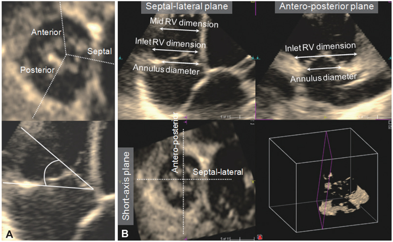
Fig. 1 Geometric measurements using real-time 3-dimensional echocardiography. A: three longitudinal planes that perpendicularly cross the middle of each leaflet are generated by guidance on the short axis image of the tricuspid valve (upper panel). On each of these three longitudinal planes, the leaflet tenting angle between the tricuspid annulus line and the leaflet is measured on a mid-systole frame (lower panel). B: mutually perpendicular septal-lateral and antero-posterior longitudinal planes are created to measure septal-lateral and antero-posterior tricuspid annulus diameters, inlet right ventricular (RV) dimensions and mid RV septal-lateral dimension.

Fig. 2 Right ventricular (RV) volume measurement using real-time 3-dimensional echocardiography. RV end-systolic and end-diastolic volumes, and ejection fraction were measured using 4-dimensional RV analysis program.
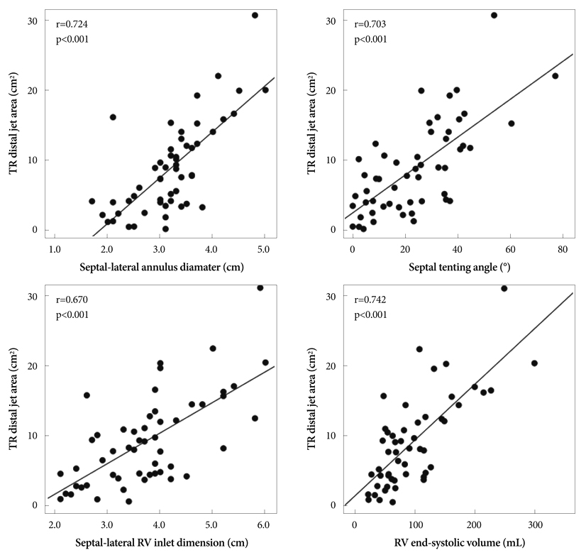
Fig. 3 Correlations between distal jet area of tricuspid regurgitation (TR) and geometric variables of tricuspid valve and right ventricle (RV).
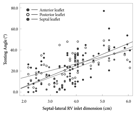
Fig. 4 Correlations between septal-lateral right ventricular (RV) inlet dimension and tenting angles of the 3 leaflets.

Fig. 5 Correlations between annulus diameters and right ventricular (RV) inlet dimensions of the corresponding directions.
Reference
-
1. Sagie A, Schwammenthal E, Padial LR, Vazquez de Prada JA, Weyman AE, Levine RA. Determinants of functional tricuspid regurgitation in incomplete tricuspid valve closure: Doppler color flow study of 109 patients. J Am Coll Cardiol. 1994. 24:446–453.2. Ton-Nu TT, Levine RA, Handschumacher MD, et al. Geometric determinants of functional tricuspid regurgitation: insights from 3-dimensional echocardiography. Circulation. 2006. 114:143–149.3. Park YH, Song JM, Lee EY, Kim YJ, Kang DH, Song JK. Geometric and hemodynamic determinants of functional tricuspid regurgitation: a real-time three-dimensional echocardiography study. Int J Cardiol. 2008. 124:160–165.4. Fukuda S, Saracino G, Matsumura Y, et al. Three-dimensional geometry of the tricuspid annulus in healthy subjects and in patients with functional tricuspid regurgitation: a real-time, 3-dimensional echocardiographic study. Circulation. 2006. 114:1 Suppl. I492–I498.5. Sukmawan R, Watanabe N, Ogasawara Y, et al. Geometric changes of tricuspid valve tenting in tricuspid regurgitation secondary to pulmonary hypertension quantified by novel system with transthoracic real-time 3-dimensional echocardiography. J Am Soc Echocardiogr. 2007. 20:470–476.6. Kanter KR, Doelling NR, Fyfe DA, Sharma S, Tam VK. De Vega tricuspid annuloplasty for tricuspid regurgitation in children. Ann Thorac Surg. 2001. 72:1344–1348.7. Bonow RO, Carabello BA, Chatterjee K, et al. 2008 Focused update incorporated into the ACC/AHA 2006 guidelines for the management of patients with valvular heart disease: a report of the American College of Cardiology/American Heart Association Task Force on Practice Guidelines (Writing Committee to Revise the 1998 Guidelines for the Management of Patients With Valvular Heart Disease): endorsed by the Society of Cardiovascular Anesthesiologists, Society for Cardiovascular Angiography and Interventions, and Society of Thoracic Surgeons. Circulation. 2008. 118:e523–e661.8. Kwon DA, Shin DH, Jung JW, et al. Echocardiographic parameters for predicting the outcome of patients undergoing surgery for severe tricuspid regurgitation. Korean Circ J. 2005. 35:916–920.9. Fukuda S, Song JM, Gillinov AM, et al. Tricuspid valve tethering predicts residual tricuspid regurgitation after tricuspid annuloplasty. Circulation. 2005. 111:975–979.10. Fukuda S, Gillinov AM, McCarthy PM, et al. Determinants of recurrent or residual functional tricuspid regurgitation after tricuspid annuloplasty. Circulation. 2006. 114:1 Suppl. I582–I587.11. Tamborini G, Brusoni D, Torres Molina JE, et al. Feasibility of a new generation three-dimensional echocardiography for right ventricular volumetric and functional measurements. Am J Cardiol. 2008. 102:499–505.12. Zoghbi WA, Enriquez-Sarano M, Foster E, et al. Recommendations for evaluation of the severity of native valvular regurgitation with two-dimensional and Doppler echocardiography. J Am Soc Echocardiogr. 2003. 16:777–802.13. Koelling TM, Aaronson KD, Cody RJ, Bach DS, Armstrong WF. Prognostic significance of mitral regurgitation and tricuspid regurgitation in patients with left ventricular systolic dysfunction. Am Heart J. 2002. 144:524–529.14. Nath J, Foster E, Heidenreich PA. Impact of tricuspid regurgitation on long-term survival. J Am Coll Cardiol. 2004. 43:405–409.15. Louie EK, Bieniarz T, Moore AM, Levitsky S. Reduced atrial contribution to left ventricular filling in patients with severe tricuspid regurgitation after tricuspid valvulectomy: a Doppler echocardiographic study. J Am Coll Cardiol. 1990. 16:1617–1624.16. Reynertson SI, Kundur R, Mullen GM, Costanzo MR, McKiernan TL, Louie EK. Asymmetry of right ventricular enlargement in response to tricuspid regurgitation. Circulation. 1999. 100:465–467.17. Mukherjee D, Nader S, Olano A, Garcia MJ, Griffin BP. Improvement in right ventricular systolic function after surgical correction of isolated tricuspid regurgitation. J Am Soc Echocardiogr. 2000. 13:650–654.18. Liel-Cohen N, Guerrero JL, Otsuji Y, et al. Design of a new surgical approach for ventricular remodeling to relieve ischemic mitral regurgitation: insights from 3-dimensional echocardiography. Circulation. 2000. 101:2756–2763.19. Hung J, Guerrero JL, Handschumacher MD, Supple G, Sullivan S, Levine RA. Reverse ventricular remodeling reduces ischemic mitral regurgitation: echo-guided device application in the beating heart. Circulation. 2002. 106:2594–2600.20. Inoue M, McCarthy PM, Popovic ZB, et al. The Coapsys device to treat functional mitral regurgitation: in vivo long-term canine study. J Thorac Cardiovasc Surg. 2004. 127:1068–1076. discussion 1076-7.
- Full Text Links
-
- Actions
-
Cited
- CITED
-
- Close
- Share
-
- Similar articles
-
- Revisit of Functional Tricuspid Regurgitation; Current Trends in the Diagnosis and Management
- Tricuspid Regurgitation in Patients with Atrial Septal Defect
- The Pathophysiology of the Transient Neonatal Tricuspid Regurgitation
- Tricuspid Valve Repair for Tricuspid Valve Insufficiency Following a Cardiac Stab Injury
- Noninvasive Evaluation of Rheumatic Tricuspid Stenosis with Doppler and 2 Dimensional Echocardiography
