J Korean Soc Spine Surg.
2011 Dec;18(4):223-229. 10.4184/jkss.2011.18.4.223.
Changes in Sagittal Spinopelvic Parameters according to Pelvic Incidence in Asymptomatic Old Korean Men
- Affiliations
-
- 1Seoul Veterans Hospital, Seoul, Korea. drortho@korea.com
- 2National Police Hospital, Seoul, Korea.
- 3Columbia University Medical Center, NY, U.S.A.
- KMID: 1448000
- DOI: http://doi.org/10.4184/jkss.2011.18.4.223
Abstract
- STUDY DESIGN: A radiographic study of normal subjects.
OBJECTIVES
To analyze sagittal spinal parameters according to the size of pelvic incidence (PI). SUMMARY OF LITERATURE REVIEW: There has been no previous study about the classification of spinopelvic parameters that has used a large cohort of asymptomatic older men with the same ethnic background as those in the current study.
MATERIALS AND METHODS
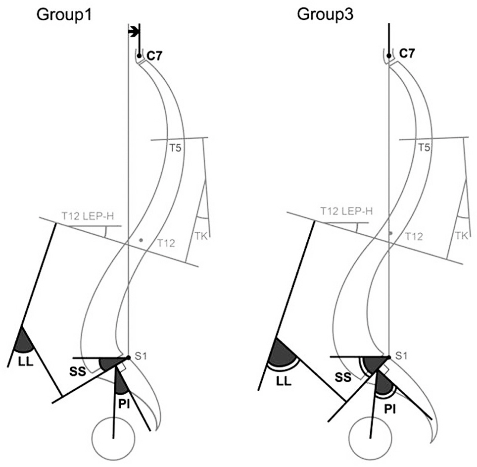
We examined 160 males aged over 50 without disease, trauma, or history of operation on spine or lower extremities. Sagittal standing radiographs of the whole spine on 36-inch film were taken. Group 1 (n=30) had a PI of less than 40degrees. Group 2 (n=71) had PI between 40degrees and 50degrees, and group 3 (n=59) had a PI greater than 50degrees. Thoracic kyphosis, thoracolumbar kyphosis, lumbar lordosis, the vertebral slope of T12, sacral slope, and pelvic incidence were measured. The distances from the plumb line of C7, T12, and the lumbar apex to the posterosuperior corner of the sacrum were also measured.
RESULTS
Subjects' average age was 64.1(53~83).Lumbar lordosis, sacral slope and pelvic tilt were all significantly increased in group 3. Thoracic kyphosis and the vertebral slope of T12 were not different between groups. The distances from the plumb line of C7, T12, and the lumbar apex to the posterosuperior corner of the sacrum were significantly translated anteriorly in group 3.
CONCLUSIONS
Group 3, who had the largest PI, demonstrated the largest lumbar lordosis and the most forward transition of trunk. However there were no differences in thoracic kyphosis and the vertebral slope of T12 among the three groups.
Keyword
MeSH Terms
Figure
Cited by 1 articles
-
A Comparative Analysis of Thoracic and Thoracolumbar Kyphosis between Young Men and Old Men
Gyu-Bok Kang, Young-Joon Ahn, Yongjung J. Kim, Youngbae B. Kim, Young-Rok Ko
J Korean Orthop Assoc. 2016;51(1):48-53. doi: 10.4055/jkoa.2016.51.1.48.
Reference
-
1. Barrey C, Jund J, Noseda O, Roussouly P. Sagittal balance of the pelvis-spine complex and lumbar degenerative diseases. A comparative study about 85 cases. Eur Spine J. 2007; 16:1459–67.
Article2. Kawakami M, Tamaki T, Ando M, Yamada H, Hashizume H, Yoshida M. Lumbar sagittal balance influences the clinical outcome after decompression and posterolateral spinal fusion for degenerative lumbar spondylolisthesis. Spine (Phila Pa 1976). 2002; 27:59–64.
Article3. Korovessis P, Dimas A, Iliopoulos P, Lambiris E. Correlative analysis of lateral vertebral radiographic variables and medical outcomes study short-form health survey: a comparative study in asymptomatic volunteers versus patients with low back pain. J Spinal Disord Tech. 2002; 15:384–90.4. Kumar MN, Baklanov A, Chopin D. Correlation between sagittal plane changes and adjacent segment degeneration following lumbar spine fusion. Eur Spine J. 2001; 10:314–9.
Article5. Videbaek T, Bunger C, Henriksen M, Egund N, Christensen FB. Sagittal Spinal Balance After Lumbar Spinal Fusion: The Impact of Anterior Column Support: Results From a Randomized Clinical Trial With an Eight- to Thirteen-Year Radiographic Followup. Spine (Phila Pa 1976). 2011; 36:183–191.6. Roussouly P, Nnadi C. Sagittal plane deformity: an overview of interpretation and management. Eur Spine J. 2010; 19:1824–36.
Article7. Duval-Beaupè re G, Schmidt C, Cosson P. A Barycentremetric study of the sagittal shape of spine and pelvis: the conditions required for an economic standing position. Ann Biomed Eng. 1992; 20:451–62.
Article8. Legaye J, Duval-Beaupè re G, Hecquet J, Marty C. Pelvic incidence: a fundamental pelvic parameter for three-dimensional regulation of spinal sagittal curves. Eur Spine J. 1998; 7:99–103.
Article9. Lee CS, Chung SS, Chung KH, Kim SR. Significance of Pelvic Incidence in the Development of Abnormal Sagittal Alignment. J Korean Orthop Assoc. 2006; 41:274–80.
Article10. Berthonnaud E, Dimnet J, Roussouly P, Labelle H. Analysis of the sagittal balance of the spine and pelvis using shape and orientation parameters. J Spinal Disord Tech. 2005; 18:40–7.
Article11. Hammerberg KW. New concepts on the pathogenesis and classification of spondylolisthesis. Spine (Phila Pa 1976). 2005; 30(6 Suppl):4–11.
Article12. Labelle H, Roussouly P, Berthonnaud , Dimnet J, O’ Brien M. The importance of spinopelvic balance in L5-s1 developmental spondylolisthesis: a review of pertinent radiologic measurements. Spine (Phila Pa 1976). 2005; 30(6 Suppl):27–34.13. Rose PS, Bridwell KH, Lenke LG, et al. Role of pelvic incidence, thoracic kyphosis, and patient factors on sagittal plane correction following pedicle subtraction osteotomy. Spine (Phila Pa 1976). 2009; 34:785–91.
Article14. Kang KB, Kim YJ, Muzaffar N, Yang JH, Kim YB, Yeo ED. Changes of Sagittal Spinopelvic Parameters in Normal Koreans with Age over 50. Asian Spine J. 2010; 4:96–101.
Article15. Hammerberg EM, Wood KB. Sagittal profile of the elderly. J Spinal Disord Tech. 2003; 16:44–50.
Article16. Kobayashi T, Atsuta Y, Matsuno T, Takeda N. A longitudinal study of congruent sagittal spinal alignment in an adult cohort. Spine (Phila Pa 1976). 2004; 29:671–6.
Article17. O'Brien MF, Kuklo TR, Blanke K, et al. Spinal Deformity Study Group Radiographic Measurement Manual. Memphis, Medtronic Sofamor Danek: Inc;2004.18. Cohen J. Statistical power analysis for the behavioral sciencesed. Lawrence Erlbaum;1988.19. Smith A, O'Sullivan P, Straker L. Classification of sagittal thoraco-lumbo-pelvic alignment of the adolescent spine in standing and its relationship to low back pain. Spine (Phila Pa 1976). 2008; 33:2101–7.
Article20. Roussouly P, Gollogly S, Berthonnaud E, Dimnet J. Classification of the normal variation in the sagittal alignment of the human lumbar spine and pelvis in the standing position. Spine (Phila Pa 1976). 2005; 30:346–53.
Article21. Ahn YJ, Kim YB, Kang KB, Lee SW, Kim YJ. Variations in Sagittal Spinopelvic Parameters According to the Lumbar Spinal Morphology in Healthy Korean Young Men. J Korean Soc Spine Surg. 2010; 17:66–73.
Article22. Boulay C, Tardieu C, Hecquet J, et al. Sagittal alignment of spine and pelvis regulated by pelvic incidence: standard values and prediction of lordosis. Eur Spine J. 2006; 15:415–22.
Article23. Vaz G, Roussouly P, Berthonnaud E, Dimnet J. Sagittal morphology and equilibrium of pelvis and spine. Eur Spine J. 2002; 11:80–7.
Article24. Lee CS, Chung SS, Kang KC, Park SJ, Shin SK. Normal patterns of Sagittal Alignment of the Spine in Young Adults Radiological analysis in a Korean population. Spine (Phila Pa 1976). 2011; 36:E1648–54.
Article25. Kim WJ, Kang JW, Yeom JS, et al. A Comparative Analysis of Sagittal Spinal Balance in 100 Asymptomatic Young and Older Aged Volunteers. J Korean Soc Spine Surg. 2003; 10:327–34.
Article26. Itoi E. Roentgenographic analysis of posture in spinal osteoporotics. Spine (Phila Pa 1976). 1991; 16:750–6.
Article27. Suzuki H, Endo K, Mizuochi J, Kobayashi H, Tanaka H, Yamamoto K. Clasped position for measurement of sagittal spinal alignment. Eur Spine J. 2010; 19:782–6.
Article28. Vialle R, Levassor N, Rillardon L, Templier A, Skalli W, Guigui P. Radiographic analysis of the sagittal alignment and balance of the spine in asymptomatic subjects. J Bone Joint Surg Am. 2005; 87:260–7.
Article29. Gelb DE, Lenke LG, Bridwell KH, Blanke K, McEnery KW. An analysis of sagittal spinal alignment in 100 asymptomatic middle and older aged volunteers. Spine (Phila Pa 1976). 1995; 20:1351–8.
Article
- Full Text Links
- Actions
-
Cited
- CITED
-
- Close
- Share
- Similar articles
-
- Changes of Spinopelvic Parameter using Iliac Screw In Surgical Correction of Sagittal Imbalance Patients
- Difference of Sagittal Spinopelvic Alignments between Degenerative Spondylolisthesis and Isthmic Spondylolisthesis
- The Sagittal Balance of the Cervical Spine: Radiographic Analysis of Interdependence between the Occipitocervical and Spinopelvic Alignment
- Variations in Sagittal Spinopelvic Parameters According to the Lumbar Spinal Morphology in Healthy Korean Young Men
- Prognostic Importance of Spinopelvic Parameters in the Assessment of Conservative Treatment in Patients with Spondylolisthesis


