Usefulness of Diffusion-Weighted MR Imaging for Breast Lesions: Comparing the Apparent Diffusion Coefficient (ADC) Values and the Pathologic Results
- Affiliations
-
- 1Department of Radiology, Soonchunhyang University Hospital, Korea. ywchang@ schmc.ac.kr
- 2Department of Preventive Medicine, Soonchunhyang University Hospital, Korea.
- KMID: 1443542
- DOI: http://doi.org/10.3348/jksr.2011.64.4.375
Abstract
- PURPOSE
We wanted to evaluate the ability of the apparent diffusion coefficient (ADC) values to differentiate between benign and malignant breast lesions and the normal breast parenchyma.
MATERIALS AND METHODS
We used breast MRI, including DWI, to obtain images of 167 breast lesions (18 benign lesions and 149 malignant lesions) of 152 women (mean age: 48.6 years, range: 24-80 years). The mean ADC values of the malignant lesions were compared to those of the benign lesions and the normal parenchyma. We compared the ADC values of IDC, DCIS and other types of breast cancer and we also compared the ADC values with the nuclear grade of IDC.
RESULTS
The mean ADC values of the malignant lesions were lower than those of the benign lesions and the normal parenchyma (p<0.001, respectively). The mean ADC value of IDC was lower than those of DCIS and other breast cancers (p<0.001, respectively). The mean ADC value of mucinous carcinoma among the other breast cancer was characteristic high compared with that of the normal parenchyma. There was no significant differentiation of the ADC values between the nuclear grades of IDC (p<0.828). The ADC threshold value of 0.98 x 10(-3) mm2/s for discriminating between malignant and benign lesion showed a specificity of 53% and a sensitivity of 100%, and the ADC threshold value of 1.33 x 10(-3) mm2/s showed a specificity of 93% and a sensitivity of 94% for discriminating between malignant and benign lesion.
CONCLUSION
The ADC value is significantly different between the different pathological results of breast lesions.
MeSH Terms
Figure
-
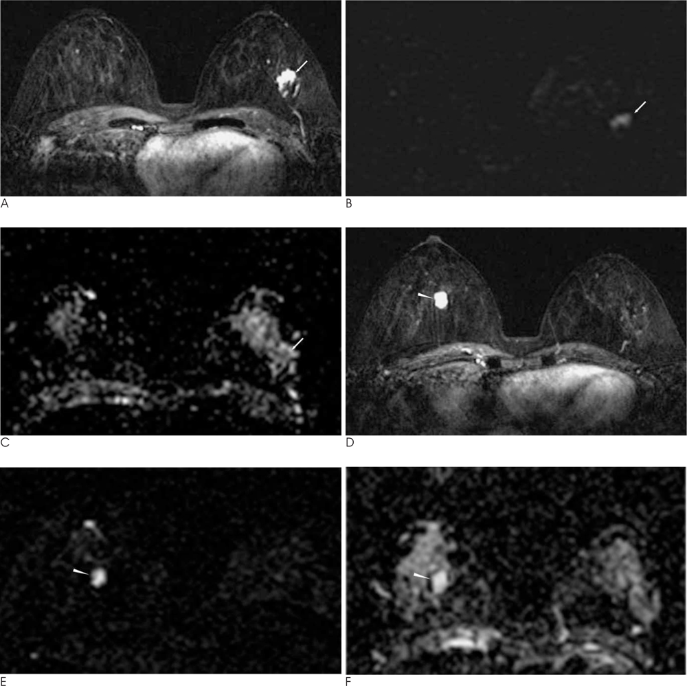
Fig. 1 45-year-old woman with infiltrative ductal carcinoma in left breast and fibroadenoma in right breast. A. Dynamic enhanced early subtraction image shows about 1.6 cm sized irregular, ill-defined, inhomogeneous enhanced mass in left upper outer quadrant (arrow). B. The diffusion weighted image shows high signal intensity lesion (arrow). C. The apparent diffusion coefficient value of the left breast tumor was 0.924 × 10⁻³mm2/s (arrow). D. Dynamic enhanced early subtraction image shows about 0.9 cm sized well-defined, round, homogeneous enhancing mass in right breast (arrowhead). E. The diffusion weighted image shows high signal intensity lesion (arrowhead). F. The apparent diffusion coefficient value of the right breast tumor was 2.062 × 10⁻³mm2/s (arrowhead).
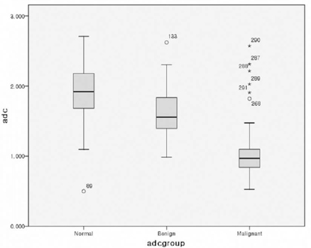
Fig. 2 Comparison among apparent diffusion coefficient values for malignant, benign and normal breast tissue.
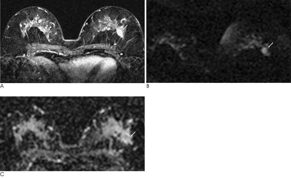
Fig. 3 65-year-old woman with ductal carcinoma in situ in left breast. A. Dynamic enhanced early subtraction image shows an ill-defined, irregular, inhomogeneously enhancing mass in left upper outer quadrant (arrow). B. The diffusion weighted image shows high signal intensity lesion (arrow). C. The apparent diffusion coefficient value of the left breast tumor was 1.302 × 10⁻³mm2/s (arrow).

Fig. 4 Comparison among apparent diffusion coefficient values of infiltrative ductal carcinoma (IDC), ductal carcinoma in situ (DCIS) and other type of malignant lesions.

Fig. 5 36-year-old woman with mucinous carcinoma in right breast. A. Dynamic enhanced early subtraction image shows an about 1.8 cm sized round, microlobulated, rim enhancing mass in right upper outer quadrant (arrow). B. The diffusion weighted image shows high signal intensity lesion (arrow). C. The apparent diffusion coefficient value of the right breast was 1.908 × 10⁻³mm2/s (arrow).
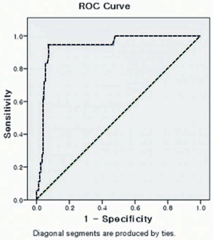
Fig. 6 Receiver operating characteristics curve for apparent diffusion coefficient value in differentiation of malignant and benign lesion. The area of under the curve is 0.936 (95% CI, 0.882-0.990, Excellent). This graph shows specificity of 53% and sensitivity of 100% for discrimination between malignant and benign breast lesion at ADC value of 0.98 × 10⁻³ mm2/s and specificity of 93% and sensitivity of 94% at ADC value of 1.33 × 10⁻³mm2/s.
Reference
-
1. Moseley ME, Cohen Y, Kucharczyk J, Mintorovitch J, Asgari HS, Wendland MF, et al. Diffusion-weighted MR imaging of anisotrophic water diffusion in cat central nervous system. Radiology. 1990; 176:439–445.2. Chenevert TL, Brunderg JA, Pipe JG. Anisotropic diffusion in human white matter: demonstration with MR techniques in vivo. Radiology. 1990; 177:401–405.3. Turner R, Le Bihan D, Maier J, Vavrek R, Hedges LK, Pekar J. Echo-planar imaging of intravoxel incoherent motion. Radiology. 1990; 177:407–414.4. Bammer R. Basic principles of diffusion-weighted imaging. Eur J Radiol. 2003; 45:169–184.5. Park MJ, Cha ES, Kang BJ, Ihn YK, Baik JH. The role of diffusionweighted imaging and the apparent diffusion coefficient (ADC) values for breast tumors. Korean J Radiol. 2007; 8:390–396.6. Woodhams R, Matsunaga K, Kan S, Hata H, Ozaki M, Iwabuchi K, et al. ADC mapping of benign and malignant breast tumor. Magn Reson Med Sci. 2005; 4:35–42.7. Hatakenaka M, Soeda H, Yabuuchi H, Matsuto Y, Kamitani T, Oda Y, et al. Apparent diffusion coefficients of breast tumors: clinical application. Magn Reson Med Sci. 2008; 7:23–29.8. Kuroki Y, Nasu K, Kuroki S, Murakami K, Hayashi T, Sekiguchi R, et al. Diffusion weighted imaging of breast cancer with the sensitivity encoding technique: analysis of the apparent diffusion coefficient value. Magn Reson Med Sci. 2004; 3:79–85.9. Woodhams R, Matsunaga K, Iwabuchi K, Kan S, Hata H, Kuranami M, et al. Diffusion-weighted imaging of malignant breast tumors: the usefulness of apparent diffusion coefficient (ADC) value and ADC map for the detection of malignant breast tumors and evaluation of cancer extension. J Comput Assist Tomogr. 2005; 29:644–649.10. Guo Y, Cai YQ, Cai ZL, Gao YG, An NY, Ma L, et al. Differentiation of clinically benign and malignant breast lesions using diffusion-weighted image. J Magn Reson Imaging. 2002; 16:172–178.11. Kawashima M, Tamaki Y, Nonaka T, Higuchi K, Kimura M, Koida T, et al. MR imaging of mucinous carcinoma of the breast. AJR Am J Roentgenol. 2002; 179:179–183.
- Full Text Links
-
- Actions
-
Cited
- CITED
-
- Close
- Share
-
- Similar articles
-
- Reversal of a Large Ischemic Lesion with Low Apparent Diffusion Coefficient Value by Rapid Spontaneous Recanalization
- Clinical applications and characteristics of apparent diffusion coefficient maps for the brain of two dogs
- Usefulness of Apparent Diffusion Coefficient in Ovarian Cystic Tumors Using Diffusion-Weighted Magnetic Resonance Imaging
- Diffusion-weighted Imaging and Apparent Diffusion Coefficient Maps for the Evaluation of Pyogenic Ventriculitis
- Intra-Individual, Inter-Vendor Comparison of Diffusion-Weighted MR Imaging of Upper Abdominal Organs at 3.0 Tesla with an Emphasis on the Value of Normalization with the Spleen
