Korean J Radiol.
2012 Oct;13(5):536-540. 10.3348/kjr.2012.13.5.536.
Trends of CT Use in the Adult Emergency Department in a Tertiary Academic Hospital of Korea during 2001-2010
- Affiliations
-
- 1Department of Radiology, Gachon University Gil Hospital, Incheon 405-760, Korea. oneshot0229@gmail.com
- 2Department of Emergency Medicine, Gachon University Gil Hospital, Incheon 405-760, Korea.
- KMID: 1392930
- DOI: http://doi.org/10.3348/kjr.2012.13.5.536
Abstract
OBJECTIVE
We wanted to assess the trends of CT examinations that were conducted in an adult emergency department (ED).
MATERIALS AND METHODS
We searched the medical database to identify adult patients (> or = 18 years) who had visited the ED and the number of CT examinations of the patients during the period from January 2001 to December 2010. We also analyzed the types of CT scans performed in terms of body parts, they were as follows; head CTs, facial bone CTs, neckl CTs, chest CTs, abdominal CTs, and miscellaneous CTs. Further, miscellaneous CTs were subdivided as CT angiography and others.
RESULTS
A total of 113656 CT scans were examined for 409439 adult ED patients during a 10-year period, and the number of CT scans increased by 255% (from 4743 CTs in 2001 to 16856 CTs in 2010), while the adult ED patient volume increased by 34% during the same period. Although the head CTs proportionally occupied the most, the facial bone CTs had the largest rate of increase (3118%), followed by cervical CTs (1173%), chest CTs (455%), miscellaneous CTs (388%; 862% and 84% for CT angiography and others, respectively), abdominal CTs (315%) and head CTs (95%) per 1000 patients during the decade.
CONCLUSION
CT use in adult ED has increased at a rate that far exceeds the growth of ED patient volume, with facial bone CTs and cervical CTs having the largest increasing rate, followed by chest CTs, miscellaneous CTs, abdominal CTs and head CTs.
Keyword
MeSH Terms
Figure
-
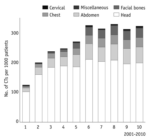
Fig. 1 Total number of CT scans performed for adult patients who visited emergency department for previous decade. CT scans per every 1000 patients increased from 124 CTs in 2001 to 330 CTs in 2010.
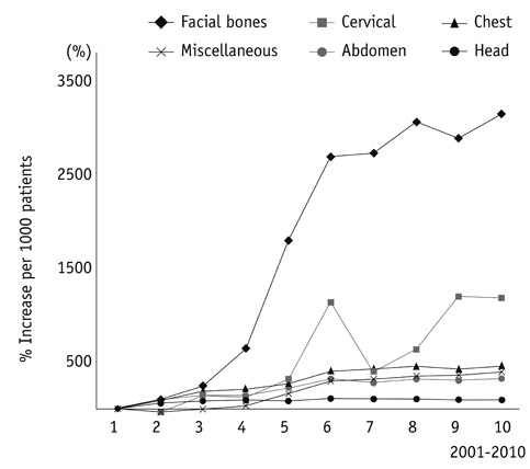
Fig. 2 Increase rate of CT examinations by anatomic region for previous decade. Compared with 2001 years, facial bone CTs shows largest rate of increase (3118%), followed by cervical CTs (1173%), chest CTs (455%), and miscellaneous CTs (388%; 862% and 84% for CT angiographies and et cetera, respectively), abdominal CTs (315%), and head CTs (95%) per 1000 patients.
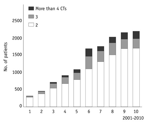
Fig. 3 Number of patients who had multiple CT examinations.
Reference
-
1. Brody AS, Seidel FG, Kuhn JP. CT evaluation of blunt abdominal trauma in children: comparison of ultrafast and conventional CT. AJR Am J Roentgenol. 1989. 153:803–806.2. Broder J, Warshauer DM. Increasing utilization of computed tomography in the adult emergency department, 2000-2005. Emerg Radiol. 2006. 13:25–30.3. Boone JM, Brunberg JA. Computed tomography use in a tertiary care university hospital. J Am Coll Radiol. 2008. 5:132–138.4. Kaewlai R, Avery LL, Asrani AV, Novelline RA. Multidetector CT of blunt thoracic trauma. Radiographics. 2008. 28:1555–1570.5. Lee J, Kirschner J, Pawa S, Wiener DE, Newman DH, Shah K. Computed tomography use in the adult emergency department of an academic urban hospital from 2001 to 2007. Ann Emerg Med. 2010. 56:591–596.6. Tanrikulu R, Erol B. Comparison of computed tomography with conventional radiography for midfacial fractures. Dentomaxillofac Radiol. 2001. 30:141–146.7. Reuben AD, Watt-Smith SR, Dobson D, Golding SJ. A comparative study of evaluation of radiographs, CT and 3D reformatted CT in facial trauma: what is the role of 3D? Br J Radiol. 2005. 78:198–201.8. Bailitz J, Starr F, Beecroft M, Bankoff J, Roberts R, Bokhari F, et al. CT should replace three-view radiographs as the initial screening test in patients at high, moderate, and low risk for blunt cervical spine injury: a prospective comparison. J Trauma. 2009. 66:1605–1609.9. Flohr TG, Schaller S, Stierstorfer K, Bruder H, Ohnesorge BM, Schoepf UJ. Multi-detector row CT systems and image-reconstruction techniques. Radiology. 2005. 235:756–773.10. Baker LC, Atlas SW, Afendulis CC. Expanded use of imaging technology and the challenge of measuring value. Health Aff (Millwood). 2008. 27:1467–1478.11. Larson DB, Johnson LW, Schnell BM, Salisbury SR, Forman HP. National trends in CT use in the emergency department: 1995-2007. Radiology. 2011. 258:164–173.12. Kock MC, Adriaensen ME, Pattynama PM, van Sambeek MR, van Urk H, Stijnen T, et al. DSA versus multi-detector row CT angiography in peripheral arterial disease: randomized controlled trial. Radiology. 2005. 237:727–737.13. Smith-Bindman R, Lipson J, Marcus R, Kim KP, Mahesh M, Gould R, et al. Radiation dose associated with common computed tomography examinations and the associated lifetime attributable risk of cancer. Arch Intern Med. 2009. 169:2078–2086.14. Brenner DJ, Elliston CD. Estimated radiation risks potentially associated with full-body CT screening. Radiology. 2004. 232:735–738.15. Sodickson A, Baeyens PF, Andriole KP, Prevedello LM, Nawfel RD, Hanson R, et al. Recurrent CT, cumulative radiation exposure, and associated radiation-induced cancer risks from CT of adults. Radiology. 2009. 251:175–184.16. Levin DC, Rao VM, Parker L. Physician orders contribute to high-tech imaging slowdown. Health Aff (Millwood). 2010. 29:189–195.17. Levin DC, Bree RL, Rao VM, Johnson J. A prior authorization program of a radiology benefits management company and how it has affected utilization of advanced diagnostic imaging. J Am Coll Radiol. 2010. 7:33–38.
- Full Text Links
-
- Actions
-
Cited
- CITED
-
- Close
- Share
-
- Similar articles
-
- Trends of CT Use in the Pediatric Emergency Department in a Tertiary Academic Hospital of Korea during 2001-2010
- The difference of Use of CT in the general versus pediatric emergency departments for adolescent patients in the same tertiary hospital
- Trends in Isolation and Antimicrobial Susceptibility of Enteropathogenic Bacteria in 2001-2010 at a Korean Tertiary Care Hospital
- An Analysis of Ten Year Trends of Cancer Incidence and Quality Control of Cancer Registration Data in Jeollabuk-do, Korea: 2001~2010
- Long-term changes in computed tomography and ultrasound utilization in a pediatric emergency department
