Canine model of ischemic stroke with permanent middle cerebral artery occlusion: clinical and histopathological findings
- Affiliations
-
- 11Department of Veterinary Internal Medicine, College of Veterinary Medicine, Konkuk University, Seoul 143-701, Korea. parkhee@konkuk.ac.kr
- 2Department of Anatomy, College of Veterinary Medicine, Konkuk University, Seoul 143-701, Korea.
- 3Acupuncture & Meridian Science Research Center, Kyunghee University, Yongin 446-701, Korea.
- 4College of Electronics and Information, Kyunghee University, Yongin 446-701, Korea.
- 5Neuroscience Research Institute, Gachon University of Medicine and Science, Incheon 405-760, Korea.
- KMID: 1106217
- DOI: http://doi.org/10.4142/jvs.2007.8.4.369
Abstract
- The aim of the present study was to assess the clinical and histopathological findings in a canine model of ischemic stroke. Cerebral ischemic stroke was induced by middle cerebral artery occlusion in four healthy beagle dogs using silicone plugs. They showed neurological signs of forebrain dysfunction such as reduced responsiveness, head turning, circling, postural reaction deficits, perceptual deficits, and hemianopsia. These signs gradually regressed within 4 weeks without therapy. On magnetic resonance imaging, T2 hyperintensity and T1 hypointensity were found in the cerebral cortex and basal ganglia. These lesions were well-defined and sharply demarcated from adjacent brain parenchyma with a homogenous appearance. No abnormalities of the cerebrospinal fluid were observed. At necropsy, atrophic and necrotic lesions were observed in the cerebral cortex. The cerebral cortex, basal ganglia, and thalamus were partially unstained with triphenyl-tetrazolium chloride. Histopathologically, typical features of infarction were identified in cortical and thalamic lesions. This study demonstrates that our canine model resembles the conditions of real stroke patients.
Keyword
MeSH Terms
Figure
-

Fig. 1 Transverse (A and B) and dorsal (C and D) T1-weighted and T2-weighted MR images of the brain in an experimentally embolized dog (ID 2). Hypointense (A and C) and hyperintense (B and D) lesions were found in lateral cortex (arrows) and caudate nucleus (arrow heads). In T2-weighted image, the well defined lesion was sharply demarcated from adjacent brain parenchyma with a homogenous appearance. Swelling, midline shift, and suppressed left lateral ventricle and thalamus by mass effect were identified in all images.
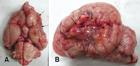
Fig. 2 The ventral (A) and lateral (B) surface of the brain (ID 2) 4 months after MCAO by a silicone embolus exhibits remarkable atrophy and necrosis (arrows) in the affected lateral cortex.
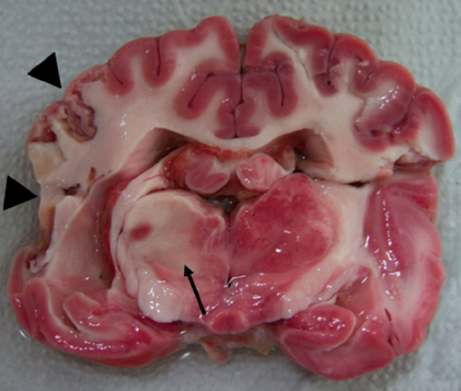
Fig. 3 Coronal section of the brain (ID 2) after TTC staining demonstrate unstained lesion on thalamus (arrow) and atrophic changes (arrow heads) on the left lateral cortex.
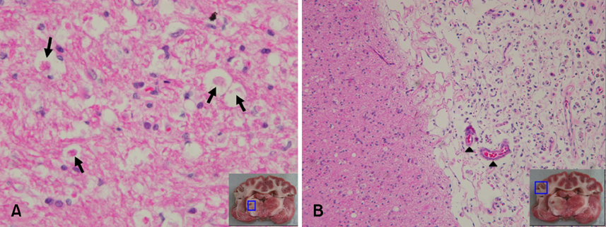
Fig. 4 Microscopic features of the brain in an experimentally embolized dog (ID 2). (A) Thalamic lesion. Necrotic neurons (arrows), nuclear pyknosis, eosinophilia of the cytoplasm, and karyolysis were prominent. H&E stain, ×400. (B) Cortex lesion. Loss of tissue cohesion, infiltration by leukocytes (especially polymorphonuclear leukocytes), congestion of small parenchymal blood vessels (arrow heads), and angioblastic proliferation were observed. H&E stain, ×100.
Cited by 1 articles
-
Canine model of ischemic stroke with permanent middle cerebral artery occlusion: clinical features, magnetic resonance imaging, histopathology, and immunohistochemistry
Joon-Hyeok Jeon, Hae-Won Jung, Hyo-Mi Jang, Jong-Hyun Moon, Ki-Tae Park, Hee-Chun Lee, Ha-Young Lim, Jung-Hyang Sur, Byeong-Teck Kang, Jeongim Ha, Dong-In Jung
J Vet Sci. 2015;16(1):75-85. doi: 10.4142/jvs.2015.16.1.75.
Reference
-
1. Berg JM, Joseph RJ. Cerebellar infarcts in two dogs diagnosed with magnetic resonance imaging. J Am Anim Hosp Assoc. 2003. 39:203–207.
Article2. Corbett RJ, Purdy PD, Laptook AR, Chaney C, Garcia D. Noninvasive measurement of brain temperature after stroke. AJNR Am J Neuroradiol. 1999. 20:1851–1857.3. D'Arceuil HE, Duggan M, He J, Pryor J, de Crespigny A. Middle cerebral artery occlusion in Macaca fascicularis: acute and chronic stroke evolution. J Med Primatol. 2006. 35:78–86.4. Diaz FG, Mastri AR, Ausman JI, Chou SN. Acute cerebral revascularization: Part I. Cerebral ischemia experimental animal model. Surg Neurol. 1979. 12:353–362.5. Garosi LS, McConnell JF. Ischaemic stroke in dogs and humans: a comparative review. J Small Anim Pract. 2005. 46:521–529.
Article6. Garosi LS, McConnell JF, Platt SR, Barone G, Baron JC, de Lahunta A, Schatzberg SJ. Results of diagnostic investigations and long-term outcome of 33 dogs with brain infarction (2000-2004). J Vet Intern Med. 2005. 19:725–731.
Article7. Garosi LS, McConnell JF, Platt SR, Barone G, Baron JC, de Lahunta A, Schatzberg SJ. Clinical and topographic magnetic resonance characteristics of suspected brain infarction in 40 dogs. J Vet Intern Med. 2006. 20:311–321.
Article8. Guo J, Liao JJ, Preston JK, Batjer HH. A canine model of acute hindbrain ischemia and reperfusion. Neurosurgery. 1995. 36:986–992.
Article9. Hillock SM, Dewey CW, Stefanacci JD, Fondacaro JV. Vascular encephalopathies in dogs: incidence, risk factors, pathophysiology, and clinical signs. Compend Contin Educ Pract Vet. 2006. 28:196–207.10. Hillock SM, Dewey CW, Stefanacci JD, Fondacaro JV. Vascular encephalopathies in dogs: diagnosis, treatment, and prognosis. Compend Contin Educ Pract Vet. 2006. 28:208–217.11. Joseph RJ, Greenlee PG, Carrillo JM, Kay WJ. Canine cerebrovascular disease: clinical and pathological findings in 17 cases. J Am Anim Hosp Assoc. 1988. 24:569–576.12. Kuwabara S, Uno J, Ishikawa S. A new model of brainstem ischemia in dogs. Stroke. 1988. 19:365–371.
Article13. McConnell JF, Garosi L, Platt SR. Magnetic resonance imaging findings of presumed cerebellar cerebrovascular accident in twelve dogs. Vet Radiol Ultrasound. 2005. 46:1–10.
Article14. Molinari GF. Experimental cerebral infarction. I. Selective segmental occlusion of intracranial arteries in the dog. Stroke. 1970. 1:224–231.
Article15. Okada Y, Shima T, Yokoyama N, Uozumi T. Comparison of middle cerebral artery trunk occlusion by silicone cylinder embolization and by trapping. J Neurosurg. 1983. 58:492–499.
Article16. Panarello GL, Dewey CW, Barone G, Stefanacci JD. Magnetic resonance imaging of two suspected cases of global brain ischemia. J Vet Emerg Crit Car. 2004. 14:269–277.
Article17. Platt SR, Garosi L. Canine cerebrovascular disease: Do dogs have strokes? J Am Anim Hosp Assoc. 2003. 39:337–342.
Article18. Purdy PD, Devous MD, Batjer HH, White CL, Meyer Y, Samson DS. Microfibrillar collagen model of canine cerebral infarction. Stroke. 1989. 20:1361–1367.
Article19. Purdy PD, Devous MD, White CL, Batjer HH, Samson DS, Brewer K, Hodges K. Reversible middle cerebral artery embolization in dogs without intracranial surgery. Stroke. 1989. 20:1368–1376.
Article20. Qureshi AI, Boulos AS, Hanel RA, Suri MFK, Yahia AM, Alberico RA, Hopkins LN. Randomized comparison of intra-arterial and intravenous thrombolysis in a canine model of acute basilar artery thrombosis. Neuroradiology. 2004. 46:988–995.
Article21. Swayne DE, Tyler DE, Batker J. Cerebral infarction with associated venous thrombosis in a dog. Vet Pathol. 1988. 25:317–320.
Article22. Thomas WB. Cerebrovascular disease. Vet Clin North Am Small Anim Pract. 1996. 26:925–943.
Article23. Traystman RJ. Animal models of focal and global cerebral ischemia. ILAR J. 2003. 44:85–95.
Article24. Yoshimoto T, Sakamoto T, Suzuki J. Experimental cerebral infarction. Part 1: Production of thalamic infarction in dogs. Stroke. 1978. 9:211–214.
Article25. Zausinger S, Schöller K, Plesnila N, Schmid-Elsaesser R. Combination drug therapy and mild hypothermia after transient focal cerebral ischemia in rats. Stroke. 2003. 34:2246–2251.
Article
- Full Text Links
-
- Actions
-
Cited
- CITED
-
- Close
- Share
-
- Similar articles
-
- Increased Expressions of eNBC and NHE1 in Ischemic Penumbra after Permanent Middle Cerebral Artery Occlusion
- Intracranial Cerebrovascular Revascularization(Extracranial-Intracranial Arterial Bypass, EIAB)
- Animal Model of Cerebral Ischemia
- ST A-MCA Anastomosis in Ischemic Stroke
- Lysophosphatidic Acid Receptor 1 Plays a Pathogenic Role in Permanent Brain Ischemic Stroke by Modulating Neuroinflammatory Responses
