Concurrent Langerhans Cell Histiocytosis and B-Lineage Lymphoid Proliferation in the Bone Marrow
- Affiliations
-
- 1Department of Laboratory Medicine, Seoul National University College of Medicine, Seoul, Korea. soonlee@snu.ac.kr
- 2Division of Hematology/Oncology, Department of Pediatrics, Seoul National University College of Medicine, Seoul, Korea.
- 3Cancer Research Institute, Seoul National University College of Medicine, Seoul, Korea.
- KMID: 1077415
- DOI: http://doi.org/10.3343/kjlm.2009.29.5.402
Abstract
- We present three cases of concurrent Langerhans cell histiocytosis (LCH) and B-lineage lymphoid cell infiltrations and/or nodules in the bone marrow. The first patient was a 25-month-old boy who presented with LCH on the right shoulder and multiple osteolytic lesions. Bone marrow biopsy showed the presence of LCH and two large lymphoid nodules of B-lineage, which were located in the paratrabecular region. Both LCH and the lymphoid nodules resolved after treatment with prednisone, vinblastine, methotrexate, and cyclophosphamide. The second patient was a 7-month-old girl who presented with LCH in the scalp and bone marrow. In spite of the treatment, a follow-up bone marrow analysis performed after 16 months showed LCH and increased B-lineage lymphoid cells in the interstitial area. The third patient was a 26-month-old girl, and imaging studies revealed reddish skin lesions and multiple osteolytic lesions. Skin biopsy and bone marrow biopsy did not show the presence of LCH; however, we initiated the treatment on the basis of the results of imaging studies. The follow-up study after 6 months showed the presence of LCH and large, patchy infiltration of B-lymphoid cells. We report three rare cases of concurrent bone marrow involvement of LCH and B-lineage lymphoid proliferation, which strongly suggest lymphoid malignancy. Further, clonal changes should be studied to elucidate the common pathogenic mechanism between the two diseases.
MeSH Terms
-
Antineoplastic Agents/therapeutic use
B-Lymphocytes/immunology/*pathology
Bone Marrow/immunology/*pathology
Cell Proliferation
Child
Child, Preschool
Cyclophosphamide/therapeutic use
Drug Therapy, Combination
Female
Histiocytosis, Langerhans-Cell/*diagnosis/drug therapy/pathology
Humans
Male
Methotrexate/therapeutic use
Prednisone/therapeutic use
Vinblastine/therapeutic use
Figure
-
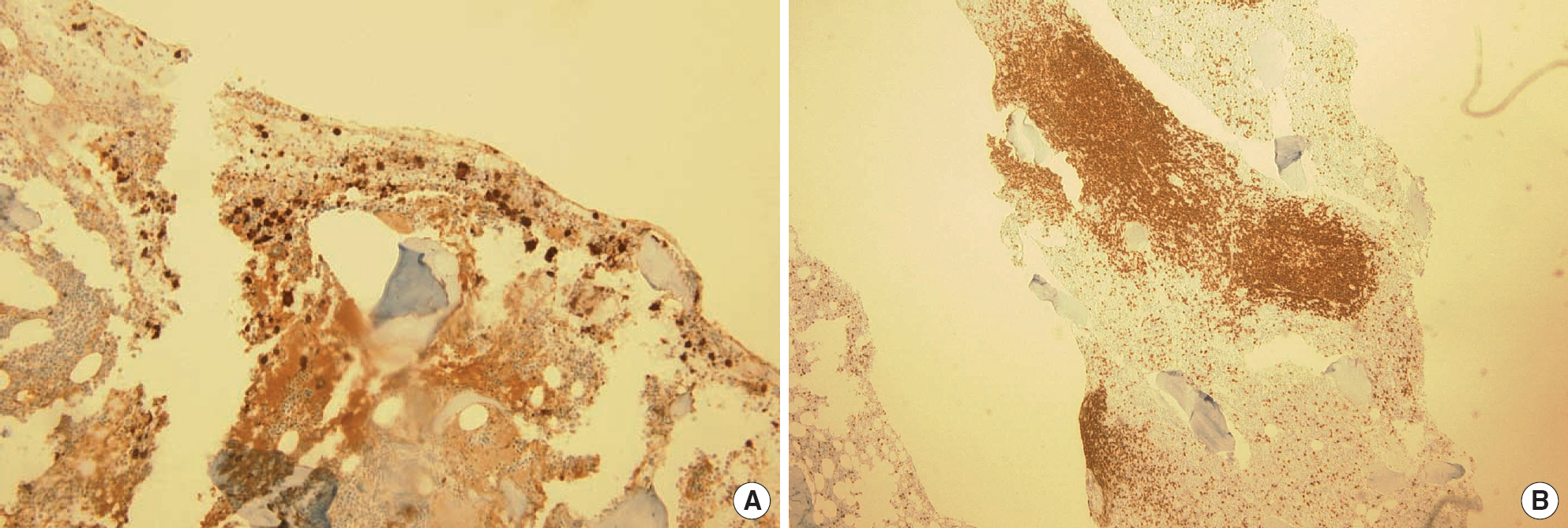
Fig. 1. Langerhans cell histiocytosis and multiple large cluster of differentiation 20 (CD20)-positive, B-lineage lymphoid nodules in the bone marrow of patient 1. (A) The left bone marrow biopsy section shows diffuse S100-positive epithelioid infiltration (×100). (B) The right bone marrow biopsy section shows two extremely large lymphoid nodules and three large CD20-positive nodules (×40).

Fig. 2. Langerhans cell histiocytosis and interstitially scattered cluster of differentiation 20 (CD20)-positive B-lymphoid cells in the bone marrow of patient 2. (A) The left bone marrow biopsy section showed increased number of S100-positive cells (× 200). (B) The right bone marrow biopsy section showed interstitial infiltration of CD20-positive cells (× 200).

Fig. 3. Langerhans cell histiocytosis and cluster of differentiation 20 (CD20)-positive B-lymphoid cell infiltration in the bone marrow of patient 3. (A) The left bone marrow biopsy section showed increased number of S100-positive cells (×200). (B) The left bone marrow biopsy section showed large, patchy infiltration of CD20-positive cells (×100).
Reference
-
1.Adu-Poku K., Thomas DW., Khan MK., Holgate CS., Smith ME. Langerhans cell histiocytosis in sequential discordant lymphoma. J Clin Pathol. 2005. 58:104–6.
Article2.Swerdlow SH., Campo E., Harris NL., Jaffe ES., Pileri SA., Stein H, et al. WHO classification of tumors of hematopoietic and lymphoid tissues. 2nd ed.Lyon, France: IARC Press;2008. p. 358–60.3.Feldman AL., Berthold F., Arceci RJ., Abramowsky C., Shehata BM., Mann KP, et al. Clonal relationship between precursor T-lymphoblastic leukemia/lymphoma and Langerhans-cell histiocytosis. Lancet Oncol. 2005. 6:435–7.4.Licci S., Boscaino A., De Palma M., Del Nonno F., D'Antonio A. Concurrence of marginal zone B-cell lymphoma MALT-type and Langerhans cell histiocytosis in a thyroid gland with Hashimoto disease. Ann Hematol. 2008. 87:855–7.
Article5.Thiele J., Zirbes TK., Kvasnicka HM., Fischer R. Focal lymphoid aggregates (nodules) in bone marrow biopsies: differentiation between benign hyperplasia and malignant lymphoma—a practical guideline. J Clin Pathol. 1999. 52:294–300.
Article
- Full Text Links
-
- Actions
-
Cited
- CITED
-
- Close
- Share
-
- Similar articles
-
- A Case of an Adult-Onset Langerhans Cell Histiocytosis Involving the Temporal Bone
- Single System Langerhans' Cell Histiocytosis with Multifocal Bone Lesions and Pathologic Fracture: A Case Report
- A Case of Orbital Langerhans' cell histiocytosis
- Fine Needle Aspiration Cytology of Langerhans' Cell Histiocytosis in the Lymph Node
- A Case of External Auditory Canal Stenosis in Langerhans Cell Histiocytosis
