J Vet Sci.
2011 Sep;12(3):209-214. 10.4142/jvs.2011.12.3.209.
Multidetector computed tomographic angiography evaluation of micropig major systemic vessels for xenotransplantation
- Affiliations
-
- 1College of Veterinary Medicine, Biotherapy Human Resources Center, Chonnam National University, Gwangju 500-757, Korea. hjhan@chonnam.ac.kr
- 2Department of Radiology, Chonnam National University Hospital, Chonnam National University Medical School, Gwangju 501-757, Korea.
- KMID: 1067391
- DOI: http://doi.org/10.4142/jvs.2011.12.3.209
Abstract
- Due primarily to the increasing shortage of allogeneic donor organs, xenotransplantation has become the focus of a growing field of research. Currently, micropigs are the most suitable donor animal for humans. However, no standard method has been developed to evaluate the systemic vascular anatomy of micropigs and standard reference values to aid in the selection of normal healthy animals as potential organ donors are lacking. Using 64-channel multidetector row computed tomographic angiography (MDCTA), we evaluated morphological features of the major systemic vessels in micropigs and compared our results to published human data. The main vasculature of the animals was similar to that of humans, except for the iliac arterial system. However, diameters of the major systemic vessels were significantly different between micropigs and humans. Specifically, the diameter of the aortic arch, abdominal aorta, external iliac artery, and femoral artery, were measured as 1.50 +/- 0.07 cm, 0.85 +/- 0.06 cm, 0.52 +/- 0.05 cm, and 0.48 +/- 0.05 cm, respectively, in the micropigs. This MDCTA data for micropig major systemic vessels can be used as standard reference values for xenotransplantation studies. The use of 64-channel MDCTA enables accurate evaluation of the major systemic vasculature in micropigs.
Keyword
MeSH Terms
Figure
-

Fig. 1 Volume-rendering image showing both the right and left common carotid arteries.
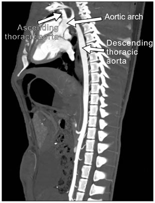
Fig. 2 Coronal maximum intensity projection showing the normal structure of the aortic arch including the ascending/descending thoracic aorta.
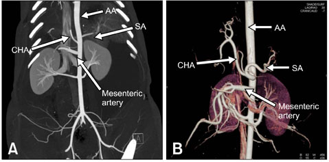
Fig. 3 Post-anterior views of the coronal maximum intensity projection (A) and the volume-rendered image (B) showing abdominal artery in the micropig. AA: abdominal aorta, CHA: common hepatic artery, SA: splenic artery.
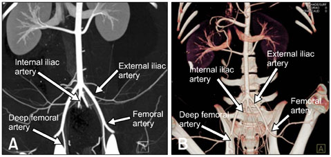
Fig. 4 Coronal maximum intensity projection (A) and 3D-CTA images (B) of the pelvic region vascular system.
Reference
-
1. Appel JZ 3rd, Buhler L, Cooper DKC. The pig as a source of cardiac xenografts. J Card Surg. 2001. 16:345–356.
Article2. Ciccone MM, Favale S, Bhuva A, Scicchitano P, Caragnano V, Lavopa C, De Pergola G, Loverro G. Anteroposterior diameter of the infrarenal abdominal aorta is higher in women with polycystic ovary syndrome. Vasc Health Risk Manag. 2009. 5:561–566.3. Cooper DKC, Gollackner B, Sachs DH. Will the pig solve the transplantation backlog? Annu Rev Med. 2002. 53:133–147.
Article4. Coşkun M, Kayahan EM, Özbek O, Çakır B, Dalgıç A, Haberal M. Imaging of hepatic arterial anatomy for depicting vascular variations in living related liver transplant donor candidates with multidetector computed tomography: comparison with conventional angiography. Transplant Proc. 2005. 37:1070–1073.
Article5. Cox A, Zhong R. Current advances in xenotransplantation. Hepatobiliary Pancreat Dis Int. 2005. 4:490–494.6. Diamond LE, Quinn CM, Martin MJ, Lawson J, Platt JL, Logan JS. A human CD46 transgenic pig model system for the study of discordant xenotransplantation. Transplantation. 2001. 71:132–142.
Article7. Duong PA, Ferson PF, Fuhrman CR, McCurry KR, Lacomis JM. 3D-multidetector CT angiography in the evaluation of potential donors for living donor lung transplantation. J Thorac Imaging. 2005. 20:17–23.
Article8. Ezzelarab M, Cooper DK. Reducing Gal expression on the pig organ-a retrospective review. Xenotransplantation. 2005. 12:278–285.
Article9. Foley WD. Special focus session: multidetector CT: abdominal visceral imaging. Radiographics. 2002. 22:701–719.10. Güven K, Acunaş B. Multidetector computed tomography angiography of the abdomen. Eur J Radiol. 2004. 52:44–55.
Article11. Hager A, Kaemmerer H, Rapp-Bernhardt U, Blücher S, Rapp K, Bernhardt TM, Galanski M, Hess J. Diameters of the thoracic aorta throughout life as measured with helical computed tomography. J Thorac Cardiovasc Surg. 2002. 123:1060–1066.
Article12. Hiroshige S, Shimada M, Harada N, Shiotani S, Ninomiya M, Minagawa R, Soejima Y, Suehiro T, Honda H, Hashizume M, Sugimachi K. Accurate preoperative estimation of liver-graft volumetry using three-dimensional computed tomography. Transplantation. 2003. 75:1561–1564.
Article13. Hosenpud JD, Bennett LE, Keck BM, Fiol B, Boucek MM, Novick RJ. The Registry of the International Society for Heart and Lung Transplantation: fifteenth official report-1998. J Heart Lung Transplant. 1998. 17:656–668.
Article14. Hunter P. Xeno's paradox. Why pig cells are better for tissue transplants than human cells. EMBO Rep. 2009. 10:554–557.15. Inomoto T, Nishizawa F, Sasaki H, Terajima H, Shirakata Y, Miyamoto S, Nagata I, Fujimoto M, Moriyasu F, Tanaka K, Yamaoka Y. Experiences of 120 microsurgical reconstructions of hepatic artery in living related liver transplantation. Surgery. 1996. 119:20–26.
Article16. Kahraman H, Ozaydin M, Varol E, Aslan SM, Dogan A, Altinbas A, Demir M, Gedikli O, Acar G, Ergene O. The diameters of the aorta and its major branches in patients with isolated coronary artery ectasia. Tex Heart Inst J. 2006. 33:463–468.17. Kamel IR, Kruskal JB, Warmbrand G, Goldberg SN, Pomfret EA, Raptopoulos V. Accuracy of volumetric measurements after virtual right hepatectomy in potential donors undergoing living adult liver transplantation. AJR Am J Roentgenol. 2001. 176:483–487.
Article18. Kawamoto S, Fishman EK. MDCT angiography of living laparoscopic renal donors. Abdom Imaging. 2006. 31:361–373.
Article19. Krejza J, Arkuszewski M, Kasner SE, Weigele J, Ustymowicz A, Hurst RW, Cucchiara BL, Messe SR. Carotid artery diameter in men and women and the relation to body and neck size. Stroke. 2006. 37:1103–1105.
Article20. Lai L, Kolber-Simonds D, Park KW, Cheong HT, Greenstein JL, Im GS, Samuel M, Bonk A, Rieke A, Day BN, Murphy CN, Carter DB, Hawley RJ, Prather RS. Production of α-1,3-galactosyltransferase knockout pigs by nuclear transfer cloning. Science. 2002. 295:1089–1092.
Article21. Lee MW, Lee JM, Lee JY, Kim SH, Park EA, Han JK, Kim YJ, Shin KS, Suh KS, Choi BI. Preoperative evaluation of the hepatic vascular anatomy in living liver donors: comparison of CT angiography and MR angiography. J Magn Reson Imaging. 2006. 24:1081–1087.
Article22. Lee SS, Kim TK, Byun JH, Ha HK, Kim PN, Kim AY, Lee SG, Lee MG. Hepatic arteries in potential donors for living related liver transplantation: evaluation with multi-detector row CT angiography. Radiology. 2003. 227:391–399.
Article23. Lerner LB, Henriques HF, Harris RD. Interactive 3-dimensional computerized tomography reconstruction in evaluation of the living renal donor. J Urol. 1999. 161:403–407.
Article24. Lin CH, Steinberg AP, Ramani AP, Abreu SC, Desai MM, Kaouk J, Goldfarb DA, Gill IS. Laparoscopic live donor nephrectomy in the presence of circumaortic or retroaortic left renal vein. J Urol. 2004. 171:44–46.
Article25. Machálek L, Holibková A, Tůma J, Houserková D. The size of the splenic hilus, diameter of the splenic artery and its branches in the human spleen. Acta Univ Palacki Olomuc Fac Med. 1998. 141:45–48.26. Murakami H, Nagashima H, Takahagi Y, Miyagawa S, Fujimura T, Toyomura K, Nakai R, Yamada M, Kurihara T, Shigehisa T, Okabe M, Seya T, Shirakura R, Kinoshita T. Transgenic pigs expressing human decay-accelerating factor regulated by porcine MCP gene promoter. Mol Reprod Dev. 2002. 61:302–311.
Article27. Niemann H, Verhoeyen E, Wonigeit K, Lorenz R, Hecker J, Schwinzer R, Hauser H, Kues WA, Halter R, Lemme E, Herrmann D, Winkler M, Wirth D, Paul D. Cytomegalovirus early promoter induced expression of hCD59 in porcine organs provides protection against hyperacute rejection. Transplantation. 2001. 72:1898–1906.
Article28. Ogata K, Platt JL. Cardiac xenotransplantation: future and limitations. Cardiology. 2004. 101:144–155.
Article29. Platt JF, Ellis JH, Korobkin M, Reige KA, Konnak JW, Leichtman AB. Potential renal donors: comparison of conventional imaging with helical CT. Radiology. 1996. 198:419–423.
Article30. Prokop M. General principles of MDCT. Eur J Radiol. 2003. 45:Suppl 1. S4–S10.
Article31. Rådegran G, Saltin B. Human femoral artery diameter in relation to knee extensor muscle mass, peak blood flow, and oxygen uptake. Am J Physiol Heart Circ Physiol. 2000. 278:H162–H167.32. Ramsoondar JJ, Máchaty Z, Costa C, Williams BL, Fodor WL, Bondioli KR. Production of α1,3-galactosyltransferase-knockout cloned pigs expressing human a α1,2-fucosylosyltransferase. Biol Reprod. 2003. 69:437–445.
Article33. Rankin SC, Jan W, Koffman CG. Noninvasive imaging of living related kidney donors: evaluation with CT angiography and gadolinium-enhanced MR angiography. AJR Am J Roentgenol. 2001. 177:349–355.34. Rubin GD, Alfrey EJ, Dake MD, Semba CP, Sommer FG, Kuo PC, Dafoe DC, Waskerwitz JA, Bloch DA, Jeffrey RB. Assessment of living renal donors with spiral CT. Radiology. 1995. 195:457–462.
Article35. Schroeder T, Nadalin S, Stattaus J, Debatin JF, Malagó M, Ruehm SG. Potential living liver donors: evaluation with an all-in-one protocol with multi-detector row CT. Radiology. 2002. 224:586–591.
Article36. Silveira LA, Silveira FB, Fazan VP. Arterial diameter of the celiac trunk and its branches. Anatomical study. Acta Cir Bras. 2009. 24:43–47.
Article37. Smith PA, Ratner LE, Lynch FC, Corl FM, Fishman EK. Role of CT angiography in the preoperative evaluation for laparoscopic nephrectomy. Radiographics. 1998. 18:589–601.
Article38. Tombul ST, Aki FT, Gunay M, Inci K, Hazirolan T, Karcaaltincaba M, Erkan I, Bakkaloglu A, Yasavul U, Bakkaloglu M. Preoperative evaluation of hilar vessel anatomy with 3-D computerized tomography in living kidney donors. Transplant Proc. 2008. 40:47–49.
Article39. Toprak A, Koc M, Tezcan H, Ozener IC, Akoglu E, Oktay A. Inferior vena cava diameter determines left ventricular geometry in continuous ambulatory peritoneal dialysis patients: an echocardiographic study. Nephrol Dial Transplant. 2003. 18:2128–2133.
Article40. Tunaci A, Yekeler E. Multidetector row CT of the kidneys. Eur J Radiol. 2004. 52:56–66.
Article41. Valastro M, Veroux M, Macarone M, Cappello D, Vizcarra D, Gagliano M, Di Mare M, Spataro M, Giuffrida G, Tallarita T, Magnano San Lio V, Veroux P. Multi-detector row CT scanner angiography in the evaluation of living kidney donors. Chir Ital. 2007. 59:337–341.42. Wilmut I, Schnieke AE, McWhir J, Kind AJ, Campbell KH. Viable offspring derived from fetal and adult mammalian cells. Nature. 1997. 385:810–813.
Article
- Full Text Links
-
- Actions
-
Cited
- CITED
-
- Close
- Share
-
- Similar articles
-
- Comparison of the Multidetector-row Computed Tomographic Angiography Axial and Coronal Planes' Usefulness for Detecting Thoracodorsal Artery Perforators
- Comparison of cardiac function and coronary angiography between conventional pigs and micropigs as measured by multidetector row computed tomography
- The Utility of 64 Channel Multidetector CT Angiography for Evaluating the Renal Vascular Anatomy and Possible Variations: a Pictorial Essay
- Congenital Anomalies of the Aortic Arch: Evaluation with the Use of Multidetector Computed Tomography
- Multidetector row computed tomography evaluation of the micropig kidney as a potential renal donor
