Comparison of cardiac function and coronary angiography between conventional pigs and micropigs as measured by multidetector row computed tomography
- Affiliations
-
- 1College of Veterinary Medicine, Biotherapy Human Resources Center, Chonnam National University, Gwangju 500-757, Korea. hjhan@chonnam.ac.kr
- 2The Heart Center, Chonnam National University Hospital, Gwangju 501-757, Korea.
- 3Cardiovascular Research Institute, Chonnam National University Hospital, Gwangju 501-757, Korea.
- 4Clinical Trial Center Chonnam National University Hospital, Gwangju 501-757, Korea.
- 5Department of Radiology, Chonnam National University Hospital, Gwangju 501-757, Korea.
- KMID: 1106226
- DOI: http://doi.org/10.4142/jvs.2008.9.2.121
Abstract
- Pigs are the most likely source animals for cardiac xenotransplantation. However, an appropriate method for estimating the cardiac function of micropigs had not been established. Computed tomography (CT) analysis aimed at estimating cardiac function and assessing the coronary arteries has not been carried out in micropigs. This study determined the feasibility of evaluating cardiac function in a micropig model using multidetector row computed tomography (MDCT) and compared the cardiac function values with those of conventional pigs. The mean age of the conventional pigs and micropigs was approximately 80 days and approximately 360 days, respectively. The mean body weight in the conventional pigs and micropigs was 29.70 +/- 0.73 and 34.10 +/- 0.98 kg, respectively. Cardiac MDCT detected ejection fractions of 52.93 +/- 3.10% and 59.00 +/- 5.56% and cardiac outputs of 1.46 +/- 0.64 l/min and 1.21 +/- 0.24 l/min in conventional pigs and micropigs, respectively. There were no significant differences in cardiac function between conventional pigs and micropigs in the reconstructed CT images. There were also no differences in the coronary angiographic images obtained by MDCT. It is expected that the results of this study will help improve understanding of cardiac function in micropigs. The data presented in this study suggest that MDCT is a feasible method for evaluating cardiac function in micropigs.
Keyword
MeSH Terms
Figure
-

Fig. 1 Left ventriculogram by computerized tomography (A: end-systolic phase, B: end-diastolic phase) in a conventional pig.
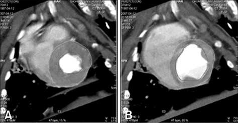
Fig. 2 Left ventriculogram by computerized tomography (A: end-systolic phase, B: end-diastolic phase) in a micropig.
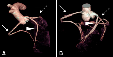
Fig. 3 Coronary circulation by computerized tomography in a conventional pig (A) and a micropig (B). Arrow; right circumflex artery (RCA). Arrowhead; left anterior descending artery (LAD). Arrow with dotted line; left circumflex artery (LCX).
Cited by 1 articles
-
Magnetic resonance imaging evaluation of Yukatan minipig brains for neurotherapy applications
Seung Pil Yun, Dong Hyun Kim, Jung Min Ryu, Jae Hong Park, Su Shin Park, Ji Hoon Jeon, Bit Na Seo, Hyun-Jeong Kim, Jun-Gyu Park, Kyoung-Oh Cho, Ho Jae Han
Lab Anim Res. 2011;27(4):309-316. doi: 10.5625/lar.2011.27.4.309.
Reference
-
1. Achenbach S, Giesler T, Ropers D, Ulzheimer S, Derlien H, Schulte C, Wenkel E, Moshage W, Bautz W, Daniel WG, Kalender WA, Baum U. Detection of coronary artery stenoses by contrast-enhanced, retrospectively electrocardiographically-gated, multislice spiral computed tomography. Circulation. 2001. 103:2535–2538.
Article2. Appel JZ 3rd, Buhler L, Cooper DK. The pig as a source of cardiac xenografts. J Card Surg. 2001. 16:345–356.
Article3. Balner H, van Leeuwen A, van Vreeswijk W, Dersjant H, van Rood JJ. Leukocytes antigens of chimpanzees and their relation to human HL-A antigens. Transplant Proc. 1970. 2:454–462.4. Barkhausen J, Ruehm SG, Goyen M, Buck T, Laub G, Debatin JF. MR evaluation of ventricular function: true fast imaging with steady-state precession versus fast low-angle shot cine MR imaging: feasibility study. Radiology. 2001. 219:264–269.
Article5. Barnes AD, Hawker RJ. Leukocyte antigens in baboons: a preliminary to tissue typing for organ grafting. Transplant Proc. 1972. 4:37–42.6. Bavelaar-Croon CDL, Kayser HWM, van der Wall EE, de Roos A, Dibbets-Schneider P, Pauwels EKJ, Germano G, Atsma DE. Left ventricular function: correlation of quantitative gated SPECT and MR imaging over a wide range of values. Radiology. 2000. 217:572–575.
Article7. Buck T, Hunold P, Wentz KU, Tkalec W, Nesser HJ, Erbel R. Tomographic three-dimensional echocardiographic determination of chamber size and systolic function in patients with left ventricular aneurysm: comparison to magnetic resonance imaging, cineventriculography, and two-dimensional echocardiography. Circulation. 1997. 96:4286–4297.
Article8. Dirksen MS, Bax JJ, de Roos A, Jukema JW, van der Geest RJ, Geleijns J, van der Wall EE, Lamb HJ. Images in cardiovascular medicine. Dynamic multislice computed tomography of left ventricular function. Circulation. 2004. 109:e25–e26.9. Dirksen MS, Bax JJ, de Roos A, Jukema JW, van der Geest RJ, Geleijns K, Boersma E, van der Wall EE, Lamb HJ. Usefulness of dynamic Multislice computed tomography of left ventricular function in unstable angina pectoris and comparison with echocardiography. Am J Cardiol. 2002. 90:1157–1160.
Article10. Fisher MR, von Schulthess GK, Higgins CB. Multiphasic cardiac magnetic resonance imaging: normal regional left ventricular wall thickening. AJR Am J Roentgenol. 1985. 145:27–30.
Article11. Germano G, Erel J, Lewin H, Kavanagh PB, Berman DS. Automatic quantitation of regional myocardial wall motion and thickening from gated technetium-99m sestamibi myocardial perfusion single-photon emission computed tomography. J Am Coll Cardiol. 1997. 30:1360–1367.
Article12. Grude M, Juergens KU, Wichter T, Paul M, Fallenberg EM, Muller JG, Heindel W, Breithardt G, Fischbach R. Evaluation of global left ventricular myocardial function with electrocardiogram-gated multidetector computed tomography: comparison with magnetic resonance imaging. Invest Radiol. 2003. 38:653–661.
Article13. Hammer C, Thein E. Determining significant physiologic incompatibilities. Graft. 2001. 4:108–110.
Article14. Hoffmann U, Moselewski F, Cury RC, Ferencik M, Jang IK, Diaz LJ, Abbara S, Brady TJ, Achenbach S. Predictive value of 16-slice multidetector spiral computed tomography to detect significant obstructive coronary artery disease in patients at high risk for coronary artery disease: patient-versus segment-based analysis. Circulation. 2004. 110:2638–2643.
Article15. Hosenpud JD, Bennett LE, Keck BM, Fiol B, Boucek MM, Novick RJ. The registry of the International Society for Heart and Lung Transplantation: fifteenth official report-1998. J heart Lung Transplant. 1998. 17:656–668.
Article16. Juergens KU, Fischbach R. Left ventricular function studied with MDCT. Eur Radiol. 2006. 16:342–357.
Article17. Juergens KU, Grude M, Maintz D, Fallenberg EM, Wichter T, Heindel W, Fischbach R. Multi-detector row CT of left ventricular function with dedicated analysis software versus MR imaging: initial experience. Radiology. 2004. 230:403–410.
Article18. Juergens KU, Maintz D, Grude M, Boese JM, Heimes B, Fallenberg EM, Heindel W, Fischbach R. Multi-detector row computed tomography of the heart: does a multi-segment reconstruction algorithm improve left ventricular volume measurements? Eur Radiol. 2005. 15:111–117.
Article19. Kirkman R. Hardy M, editor. Of swine and man: organ physiology in different species. Xenograft. 1989. vol. 25. Amsterdam: Elsevier;124–136.20. Knez A, Becker CR, Leber A, Ohnesorge B, Becker A, White C, Haberl R, Reiser MF, Steinbeck G. Usefulness of multislice spiral computed tomography angiography for determination of coronary artery stenoses. Am J Cardiol. 2001. 88:1191–1194.
Article21. Kopp AF, Schroeder S, Kuettner A, Baumbach A, Georg C, Kuzo R, Heuschmid M, Ohnesorge B, Karsch KR, Claussen CD. Non-invasive coronary angiography with high resolution multidetector-row computed tomography. Results in 102 patients. Eur Heart J. 2002. 23:1714–1725.
Article22. Lee MY, Lee SH, Lee SG, Park SH, Lee CY, Kim KH, Hwang SH, Lim SY, Ahn YK, Han HJ. Comparative analysis of heart functions in micropigs and conventional pigs using echocardiography and radiography. J Vet Sci. 2007. 8:7–14.23. Lipton MJ, Higgins CB, Farmer D, Boyd DP. Cardiac imaging with a high-speed Cine-CT Scanner: preliminary results. Radiology. 1984. 152:579–582.24. Mahnken AH, Spuentrup E, Niethammer M, Buecker A, Boese J, Wildberger JE, Flohr T, Sinha AM, Krombach GA, Gunther RW. Quantitative and qualitative assessment of left ventricular volume with ECG-gated multislice spiral CT: value of different image reconstruction algorithms in comparison to MRI. Acta Radiol. 2003. 44:604–611.
Article25. Metzgar RS, Seigler HF. Tissue antigens of man and chimpanzees; their role in xenografting. Transplant Proc. 1970. 2:463–467.26. Mochizuki T, Murase K, Higashino H, Koyama Y, Doi M, Miyagawa M, Nakata S, Shimizu K, Ikezoe J. Two- and three-dimensional CT ventriculography: a new application of helical CT. AJR Am J Roentgenol. 2000. 174:203–208.27. Nairne P, Allen I, Brazier M, Forrester D, Heap B, Kennedy I. Animal-to-human transplants: the ethics of xenotransplantation. 1996. London: Nuffield Council on Bioethics;1–123.28. Pattynama PM, Lamb HJ, van der Velde EA, van der Wall EE, de Roos A. Left ventricular measurements with cine and spin-echo MR imaging: a study of reproducibility with variance component analysis. Radiology. 1993. 187:261–268.
Article29. Qin JX, Jones M, Shiota T, Greenberg NL, Tsujino H, Firstenberg MS, Gupta PC, Zetts AD, Xu Y, Ping Sun J, Cardon LA, Odabashian JA, Flamm SD, White RD, Panza JA, Thomas JD. Validation of real-time three-dimensional echocardiography for quantifying left ventricular volumes in the presence of a left ventricular aneurysm: in vitro and in vivo studies. J Am Coll Cardiol. 2000. 36:900–907.
Article30. Raff GL, Gallagher MJ, O'Neill WW, Goldstein JA. Diagnostic accuracy of noninvasive coronary angiography using 64-slice spiral computed tomography. J Am Coll Cardiol. 2005. 46:552–557.
Article31. Ropers D, Baum U, Pohle K, Anders K, Ulzheimer S, Ohnesorge B, Schlundt C, Bautz W, Daniel WG, Achenbach S. Detection of coronary artery stenoses with thin-slice multi-detector row spiral computed tomography and multiplanar reconstruction. Circulation. 2003. 107:664–666.
Article32. Schoepf UJ, Savino G, Lake DR, Ravenel JG, Costello P. The age of CT pulmonary angiography. J Thorac Imaging. 2005. 20:273–279.33. Schuijf JD, Bax JJ, Jukema JW, Lamb HJ, Vliegen HW, Salm LP, de Roos A, van der Wall EE. Noninvasive angiography and assessment of left ventricular function using multislice computed tomography in patients with type 2 diabetes. Diabetes Care. 2004. 27:2905–2910.34. Schuijf JD, Bax JJ, Salm LP, Jukema JW, Lamb HJ, van der Wall EE, de Roos A. Noninvasive coronary imaging and assessment of left ventricular function using 16-slice computed tomography. Am J Cardiol. 2005. 95:571–574.
Article35. Stankovicova T, Szilard M, De Scheerder I, Sipido KR. M cells and transmural heterogeneity of action potential configuration in myocytes from the left ventricular wall of the pig heart. Cardiovasc Res. 2000. 45:952–960.
Article36. White HD, Norris RM, Brown MA, Brandt PW, Whitlock RM, Wild CJ. Left ventricular end-systolic volume as the major determinant of survival after recovery from myocardial infarction. Circulation. 1987. 76:44–51.
Article37. Wilmut I, Schnieke AE, McWhir J, Kind AJ, Campbell KH. Viable offspring derived from fetal and adult mammalian cells. Nature. 1997. 385:810–813.
Article38. Yamamuro M, Tadamura E, Kubo S, Toyoda H, Nishina T, Ohba M, Hosokawa R, Kimura T, Tamaki N, Komeda M, Kita T, Konishi J. Cardiac functional analysis with multi-detector row CT and segmental reconstruction algorithm: comparison with echocardiography, SPECT, and MR imaging. Radiology. 2005. 234:381–390.
- Full Text Links
-
- Actions
-
Cited
- CITED
-
- Close
- Share
-
- Similar articles
-
- Coronary Angiography with Multidetector row Computed Tomography: Part I - Technical Aspects
- A case of coronary artery-pulmonary artery fistula communicated with aorto-pulmonary fistula via common channel detected by Multidetector row CT (MDCT) and coronary angiography
- Unusual Coronary Artery Fistula: Left Anterior Descending Coronary Artery - Left Ventricular Fistula Diagnosed by ECG-Gated Multi-Detector Row Coronary CT Angiography
- Giant coronary aneurysm caused by Kawasaki disease: consistency between catheter angiography and electrocardiogram gated dual-source computed tomography angiography
- Coronary MDCT and MRI
