Comparison of Spectral-Domain and Time-Domain Optical Coherence Tomography in Solar Retinopathy
- Affiliations
-
- 1Department of Ophthalmology, Kim's Eye Hospital, Konyang University College of Medicine, Seoul, Korea. -medical-@hanmail.net
- KMID: 1018418
- DOI: http://doi.org/10.3341/kjo.2011.25.4.278
Abstract
- The purpose of this article is to compare spectral-domain (SD) and time-domain (TD) optical coherence tomography (OCT) findings in patients with solar retinopathy. Complete ocular examinations and OCT were performed in two patients presenting with acute solar retinopathy soon after observation of an eclipse. Both patients were evaluated with SD-OCT and TD-OCT at the same time. SD-OCT demonstrated characteristic defects at the level of the inner and outer segment junction of the photoreceptors in all the affected eyes and decreased reflectiveness of the retinal pigment epithelium layer. TD-OCT images showed unremarkable findings in two eyes with deteriorated visual acuity. SD-OCT improves diagnosis and assessment of the degree and nature of foveal damage in patients with solar retinopathy and may be an important tool for use in identifying foveal damage not detected by TD-OCT. SD-OCT may be preferable to TD-OCT for confirmation or assessment of the degree of foveal damage in patients with solar retinopathy.
MeSH Terms
Figure
-
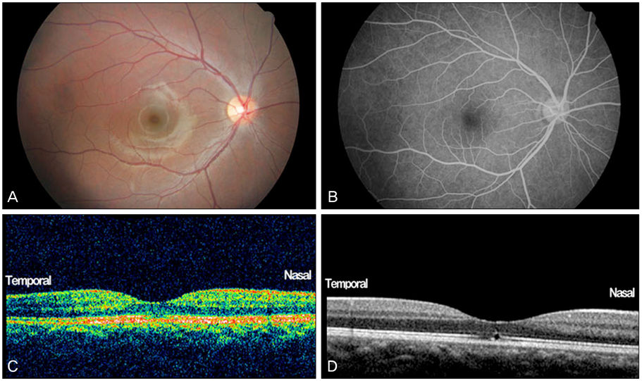
Fig. 1 (A) Fundus photograph of the right eye in case 1 showing a small hypopigmented lesion in the fovea. (B) Fluorescein angiogram of the right eye of the same patient showing unremarkable findings. (C) Time-domain optical coherence tomography (OCT) through the fovea of the right eye of the same patient showing the smallest possible abnormality of the inner hyperreflective layer. (D) Spectral-domain-OCT of the same patient confirmed more clearly the disruption of the inner segment and outer segment line, and decreased intensity of the reflectiveness of the retinal pigment epithelium at the fovea.
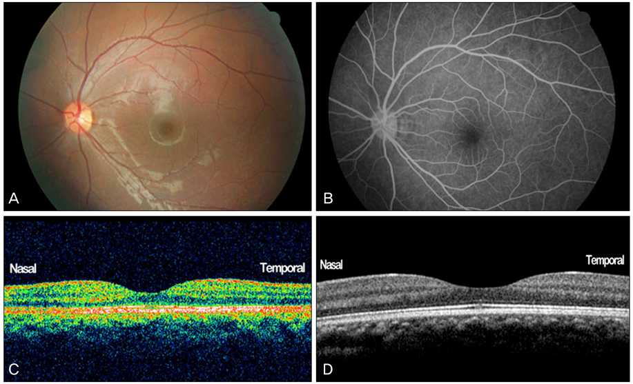
Fig. 2 (A) Fundus photograph of the left eye in case 2, showing unremarkable findings. (B) Fluorescein angiography of the same patient showing unremarkable findings. (C) Time-domain-optical coherence tomography (OCT) through the fovea of the left eye of the same patient showing unremarkable findings. (D) Spectral-domain-OCT of the same patient revealed a small disruption of the inner segment and outer segment line, and decreased intensity of the reflectiveness of the retinal pigment epithelium at the fovea.
Cited by 1 articles
-
A Case of Acute Bilateral Solar Retinopathy Diagnosed with Spectral Domain Optical Coherence Tomography
Jin Young Kwon, Jong Hyun Jung, Do Gyun Kim
J Korean Ophthalmol Soc. 2015;56(12):1974-1978. doi: 10.3341/jkos.2015.56.12.1974.
Reference
-
1. Galainena ML. Solar retinopathy. Ann Ophthalmol. 1976. 8:304–306.2. Bechmann M, Ehrt O, Thiel MJ, et al. Optical coherence tomography findings in early solar retinopathy. Br J Ophthalmol. 2000. 84:547–548.3. Jain A, Desai RU, Charalel RA, et al. Solar retinopathy: comparison of optical coherence tomography (OCT) and fluorescein angiography (FA). Retina. 2009. 29:1340–1345.4. Srinivasan VJ, Wojtkowski M, Witkin AJ, et al. High-definition and 3-dimensional imaging of macular pathologies with high-speed ultrahigh-resolution optical coherence tomography. Ophthalmology. 2006. 113:2054.e1–2054.14.5. Gass JD. Stereoscopic atlas of macular disease: diagnosis and treatment. 1987. 3rd ed. St. Louis: Mosby;570–572.6. Tso MO, La Piana FG. The human fovea after sungazing. Trans Sect Ophthalmol Am Acad Ophthalmol Otolaryngol. 1975. 79:OP788–OP795.7. Calvo-González C, Reche-Frutos J, Santos-Bueso E, et al. Optical coherence tomography in solar eclipse retinopathy. Arch Soc Esp Oftalmol. 2006. 81:297–300.
- Full Text Links
-
- Actions
-
Cited
- CITED
-
- Close
- Share
-
- Similar articles
-
- A Case of Acute Bilateral Solar Retinopathy Diagnosed with Spectral Domain Optical Coherence Tomography
- Choroidal Thickness at the Outside of Fovea in Diabetic Retinopathy Using Spectral-Domain Optical Coherence Tomography
- Comparison of the Efficacy between Time and Spectral Domain Optical Coherence Tomography for the Identification of Vitreomacular Interface
- Macular Thickness Changes with Age and Gender in Emmetropia Using Spectral Domain Optical Coherence Tomography
- Difference of GCIPL Thickness of Diabetes and Normal Eyes in Spectral Domain OCT
