Macular Gradient Measurement in Myopic Posterior Staphyloma Using Optical Coherence Tomography
- Affiliations
-
- 1Department of Ophthalmology, Chungbuk National University College of Medicine, Cheongju, Korea.
- 2Department of Ophthalmology, Asan Medical Center, University of Ulsan College of Medicine, Seoul, Korea. dropkim@dreamwiz.com
- 3Department of Ophthalmology, Gangneung Asan Medical Center, University of Ulsan College of Medicine, Gangneung, Korea.
- KMID: 1018411
- DOI: http://doi.org/10.3341/kjo.2011.25.4.243
Abstract
- PURPOSE
To evaluate clinical characteristics and the macular gradient in myopic posterior staphyloma with time domain (TD) optical coherence tomography (OCT).
METHODS
Sixty-four staphyloma eyes of 40 patients were examined. Macular gradient (tangent theta) and the location of staphyloma were assessed with OCT imaging. The macular gradient was measured at points 1 mm and 2 mm distant from the fovea. The relationships of the macular gradient with age, axial length, and spherical equivalent were analyzed.
RESULTS
In 8 eyes (12.5%), the bottoms of the staphylomas were in the fovea, and there was no macular gradient. However, in the other 56 eyes (87.5%), the bottoms of the staphylomas were not in the foveal area, and macular gradients existed. Staphylomas were commonly located in the infero-nasal retinal area. The mean macular gradient (tangent theta) was 0.26 +/- 0.08 at 1 mm distance from the fovea and 0.28 +/- 0.10 at 2 mm. No significant relationships were observed between macular gradient and axial length, patient age, or spherical equivalent.
CONCLUSIONS
TD OCT reveals staphyloma location. If the location is outside of the fovea, a macular gradient exists and can be measured by OCT. Axial length measurement error may occur in eyes with poor visual fixation and steep macular gradients.
Keyword
MeSH Terms
Figure
-
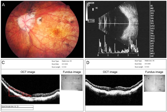
Fig. 1 Images showing posterior staphyloma. A 51-year-old woman with 20 / 40 visual acuity and a -11 diopter spherical equivalent. (A) Fundus photograph showing the posterior staphyloma. (B) On B-scan ultrasonography, a posterior staphyloma was detected. (C) An optical coherence tomography (OCT) scan showed the macular gradient. The scanning direction is transverse. Between the macula and the optic disc area, a steep macular gradient is observed, and the staphyloma protrudes toward the temporal retina. The macular gradient, between the macula and the optic disc, was measured (tangent θ). (D) On vertical scanning, symmetric posterior curvature of the retina was seen. From images of the two-axis OCT scans, the location of the staphyloma was confirmed to lie in the temporal area, with respect to the fovea.
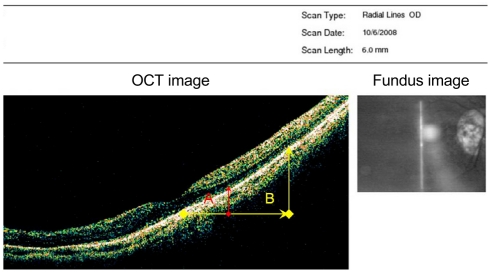
Fig. 2 Image showing a radial line scan optical coherence tomography (OCT) map. The transverse scan length (6 mm) and the vertical scan length (2 mm) are features of the Stratus OCT program. We measured the macular gradient at points 1 mm (A) and 2 mm (B) distant from the base of the fovea.
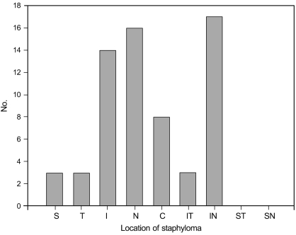
Fig. 3 The distribution of staphylomas, as shown by optical coherence tomography, in 64 eyes. Most staphylomas were distributed in the inferior and nasal retinal areas, with respect to the fovea. S = superior; T = temporal; I = inferior; N = nasal; C = center of fovea; IT = infero-temporal; IN = infero-nasal; ST = supero-temporal; SN = supero-nasal.
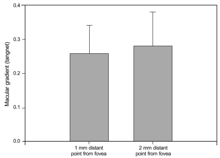
Fig. 4 Macular gradients 1 mm and 2 mm distant from the fovea. Macular gradient at 1mm was 0.26 and 0.28 at 2 mm. At 2 mm, the tangent value was greater than at 1 mm, but this was not statistically significant.
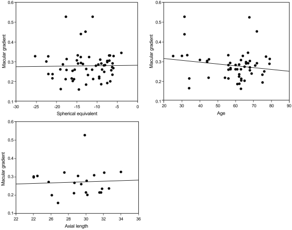
Fig. 5 Correlations between macular gradient and spherical equivalent, age, and axial length. No statistically significant associations were seen (spherical equivalent p-value, 0.728; age, 0.244, and axial length, 0.799).
Reference
-
1. Hsiang HW, Ohno-Matsui K, Shimada N, et al. Clinical characteristics of posterior staphyloma in eyes with pathologic myopia. Am J Ophthalmol. 2008; 146:102–110. PMID: 18455142.
Article2. Grossniklaus HE, Green WR. Pathologic findings in pathologic myopia. Retina. 1992; 12:127–133. PMID: 1439243.
Article3. Wong TY, Foster PJ, Hee J, et al. Prevalence and risk factors for refractive errors in adult Chinese in Singapore. Invest Ophthalmol Vis Sci. 2000; 41:2486–2494. PMID: 10937558.4. Curtin BJ. The pathogenesis of congenital myopia. A study of 66 cases. Arch Ophthalmol. 1963; 6:166–173. PMID: 14024344.5. Curtin BJ. The posterior staphyloma of pathologic myopia. Trans Am Ophthalmol Soc. 1977; 75:67–86. PMID: 613534.6. Gaucher D, Erginay A, Lecleire-Collet A, et al. Dome-shaped macula in eyes with myopic posterior staphyloma. Am J Ophthalmol. 2008; 145:909–914. PMID: 18342827.
Article7. Takano M, Kishi S. Foveal retinoschisis and retinal detachment in severely myopic eyes with posterior staphyloma. Am J Ophthalmol. 1999; 128:472–476. PMID: 10577588.
Article8. Toranzo J, Cohen SY, Erginay A, Gaudric A. Peripapillary intrachoroidal cavitation in myopia. Am J Ophthalmol. 2005; 140:731–732. PMID: 16226529.
Article9. Sayanagi K, Ikuno Y, Gomi F, Tano Y. Retinal vascular microfolds in highly myopic eyes. Am J Ophthalmol. 2005; 139:658–663. PMID: 15808161.
Article10. Nakanishi H, Tsujikawa A, Gotoh N, et al. Macular complications on the border of an inferior staphyloma associated with tilted disc syndrome. Retina. 2008; 28:1493–1501. PMID: 18667957.
Article11. Leys AM, Cohen SY. Subretinal leakage in myopic eyes with a posterior staphyloma or tilted disk syndrome. Retina. 2002; 22:659–665. PMID: 12441740.
Article12. Tsuboi S, Uchihori Y, Manabe R. Subretinal neovascularisation in eyes with localised inferior posterior staphylomas. Br J Ophthalmol. 1984; 68:869–872. PMID: 6210100.
Article13. Prost M, De Laey JJ. Choroidal neovascularization in tilted disc syndrome. Int Ophthalmol. 1988; 12:131–135. PMID: 2466011.
Article
- Full Text Links
-
- Actions
-
Cited
- CITED
-
- Close
- Share
-
- Similar articles
-
- The Shortest Radius of Curvature of Bruch's Membrane in Macular Optical Coherence Tomography
- Macular Buckling Surgery Using a Novel L-shaped Buckle for Patients with Myopic Tractional Maculopathy
- Use of Spectral-Domain Optical Coherence Tomography to Analyze Macular Thickness According to Refractive Error
- Incidence and Risk Factors of Cystoid Macular Edema after Vitrectomy with Silicone Oil Tamponade for Retinal Detachment
- The Correlation between Visual Acuity and Patterns of Diabetic Macular Edema in OCT Images
