Korean J Ophthalmol.
2011 Aug;25(4):238-242. 10.3341/kjo.2011.25.4.238.
Evaluation of Changes of Macular Thickness in Diabetic Retinopathy after Cataract Surgery
- Affiliations
-
- 1Department of Ophthalmology, Hallym University Sacred Heart Hospital, Anyang, Korea. piw@korea.com
- KMID: 1018410
- DOI: http://doi.org/10.3341/kjo.2011.25.4.238
Abstract
- PURPOSE
To assess the macular thickness changes after cataract surgery in diabetic patients using optical coherence tomography (OCT).
METHODS
We retrospectively reviewed the records of 104 diabetic patients who underwent cataract surgery. We examined the changes of macular thickness using OCT before cataract surgery and 1 week, 1-, 2- and 6-months after surgery. The central subfield mean thickness (CSMT) was used to evaluate macular edema which was defined as an increase of CSMT (DeltaCSMT) > 30% from the baseline. The association between prior laser treatment or severity of diabetic retinopathy and macular thickness were also analyzed.
RESULTS
Macular edema occurred in 19 eyes (18%) from the diabetic group and 63% of macular edema developed at 1 month after surgery. Thirteen (68%) out of 19 eyes with macular edema showed the resolution of macular edema by 6 months after surgery without treatment. DeltaCSMT of eyes without a history of laser treatment was statistically greater compared to eyes with a history of laser treatment in at 1- and 2-months after surgery, but was not different than eyes who had laser treatment at 6-months after surgery. The severity of diabetic retinopathy was not significantly correlated to macular edema, but there was statistical difference when patients who had a history of prior laser treatment were excluded.
CONCLUSIONS
The incidence of macular edema after cataract surgery in diabetic patients was 18%. Its peak incidence was at 1 month post surgery and it resolved spontaneously in 68% of patients by 6 months post surgery. Prior laser treatment might prevent postoperative macular edema until 2 months after cataract surgery in diabetic patients. However, macular edema did not affect the severity of diabetic retinopathy.
MeSH Terms
-
Aged
Cataract/*complications
*Cataract Extraction
Diabetic Retinopathy/complications/*pathology
Disease Progression
Female
Follow-Up Studies
Humans
Macula Lutea/*pathology
Macular Degeneration/etiology/pathology
Male
Postoperative Period
Prognosis
Retrospective Studies
Time Factors
Tomography, Optical Coherence
Visual Acuity
Figure
-
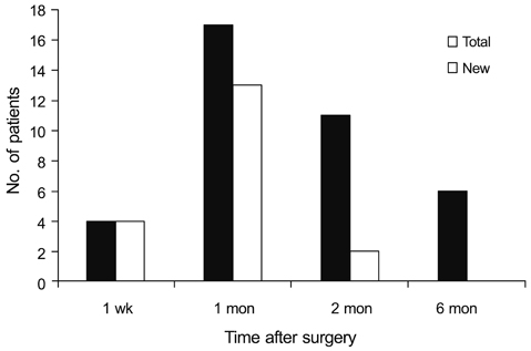
Fig. 1 The total and newly developed number of patients with macula edema after cataract surgery.
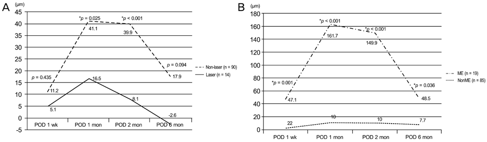
Fig. 2 The changes of central subfield macular thickness in eyes with and without prior laser treatment (A) and in eyes with and without macular edema (B) after cataract surgery. POD = postoperative day; ME = macular edema. *Student t-test (p < 0.05).
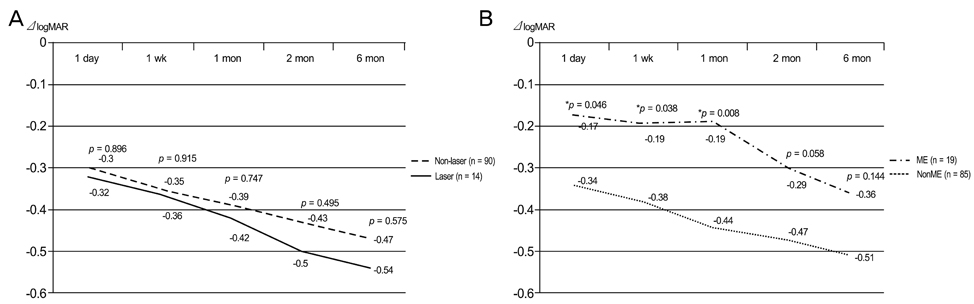
Fig. 3 The changes of best corrected visual acuity in eyes with and without prior laser treatment (A) and in eyes with and without macular edema (B) after cataract surgery. logMAR = logarithm of the minimal angle of resolution; ME = macular edema. *Student t-test (p < 0.05).
Reference
-
1. Wright PL, Wilkinson CP, Balyeat HD, et al. Angiographic cystoid macular edema after posterior chamber lens implantation. Arch Ophthalmol. 1988. 106:740–744.2. Kim SJ, Equi R, Bressler NM. Analysis of macular edema after cataract surgery in patients with diabetes using optical coherence tomography. Ophthalmology. 2007. 114:881–889.3. Hee MR, Puliafito CA, Wong C, et al. Quantitative assessment of macular edema with optical coherence tomography. Arch Ophthalmol. 1995. 113:1019–1029.4. Goebel W, Kretzchmar-Gross T. Retinal thickness in diabetic retinopathy: a study using optical coherence tomography (OCT). Retina. 2002. 22:759–767.5. Browning DJ, McOwen MD, Bowen RM Jr, O'Marah TL. Comparison of the clinical diagnosis of diabetic macular edema with diagnosis by optical coherence tomography. Ophthalmology. 2004. 111:712–715.6. Dowler JG, Sehmi KS, Hykin PG, Hamilton AM. The natural history of macular edema after cataract surgery in diabetes. Ophthalmology. 1999. 106:663–668.7. Browning DJ, Glassman AR, Aiello LP, et al. Optical coherence tomography measurements and analysis methods in optical coherence tomography studies of diabetic macular edema. Ophthalmology. 2008. 115:1366–1371. –1371.e1.8. Gass JD, Norton EW. Cystoid macular edema and papilledema following cataract extraction. A fluorescein fundoscopic and angiographic study. Arch Ophthalmol. 1966. 76:646–661.9. Benson WE. Cataract surgery and diabetic retinopathy. Curr Opin Ophthalmol. 1992. 3:396–400.10. Funatsu H, Yamashita H, Noma H, et al. Prediction of macular edema exacerbation after phacoemulsification in patients with nonproliferative diabetic retinopathy. J Cataract Refract Surg. 2002. 28:1355.11. Krepler K, Biowski R, Schrey S, et al. Cataract surgery in patients with diabetic retinopathy: visual outcome, progression of diabetic retinopathy, and incidence of diabetic macular oedema. Graefes Arch Clin Exp Ophthalmol. 2002. 240:735–738.12. Early Treatment Diabetic Retinopathy Study research group. Photocoagulation for diabetic macular edema. Early Treatment Diabetic Retinopathy Study report number 1. Arch Ophthalmol. 1985. 103:1796–1806.13. Pollack A, Leiba H, Bukelman A, Oliver M. Cystoid macular oedema following cataract extraction in patients with diabetes. Br J Ophthalmol. 1992. 76:221–224.14. Chew EY, Benson WE, Remaley NA, et al. Results after lens extraction in patients with diabetic retinopathy: early treatment diabetic retinopathy study report number 25. Arch Ophthalmol. 1999. 117:1600–1606.
- Full Text Links
-
- Actions
-
Cited
- CITED
-
- Close
- Share
-
- Similar articles
-
- The Changes in Central Macular Thickness after Cataract Surgery in Patients with Diabetic Retinopathy
- The Changes in Macular Thickness after Phacoemulsification in Patients with Non-diabetes and Nonproliferative Diabetic Retinopathy
- The Clinical Evaluation about the Impact of Cataract Operation on the Progression of Diabetic Retinopathy
- Changes in Macular Thickness after Cataract Surgery According to Optical Coherence Tomography
- Retinal Thickness After Cataract Surgery Measured by Optical Coherence Tomography
